This site needs JavaScript to work properly. Please enable it to take advantage of the complete set of features!
Skip to main page content
U.S. flag

An official website of the United States government

Dot gov

The .gov means it’s official.
Federal government websites often end in .gov or .mil. Before sharing sensitive information, make sure you’re on a federal government site.

Https

The site is secure.
The https:// ensures that you are connecting to the official website and that any information you provide is encrypted and transmitted securely.

NIH NLM Logo
Log in
Access keys NCBI Homepage MyNCBI Homepage Main Content Main Navigation
. 2019 Feb 4;9(1):1251.
doi: 10.1038/s41598-018-37398-6.

Extracellular polysaccharide synthesis in a bloom-forming strain of Microcystis aeruginosa: implications for colonization and buoyancy

Affiliations

Extracellular polysaccharide synthesis in a bloom-forming strain of Microcystis aeruginosa: implications for colonization and buoyancy

Meng Chen et al. Sci Rep. .

Abstract

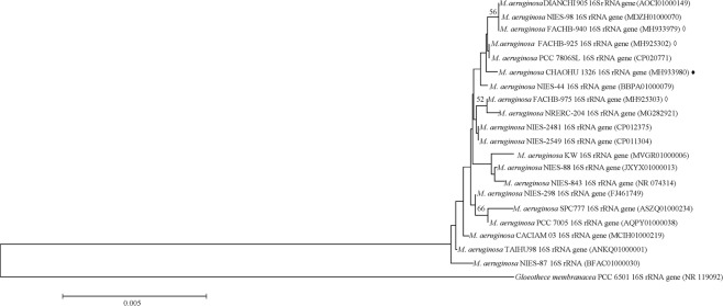
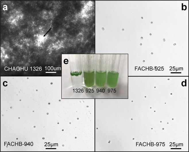
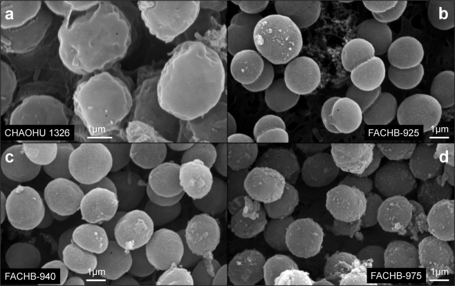
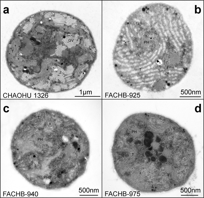
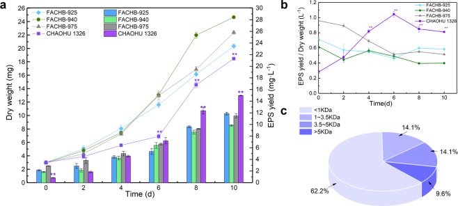
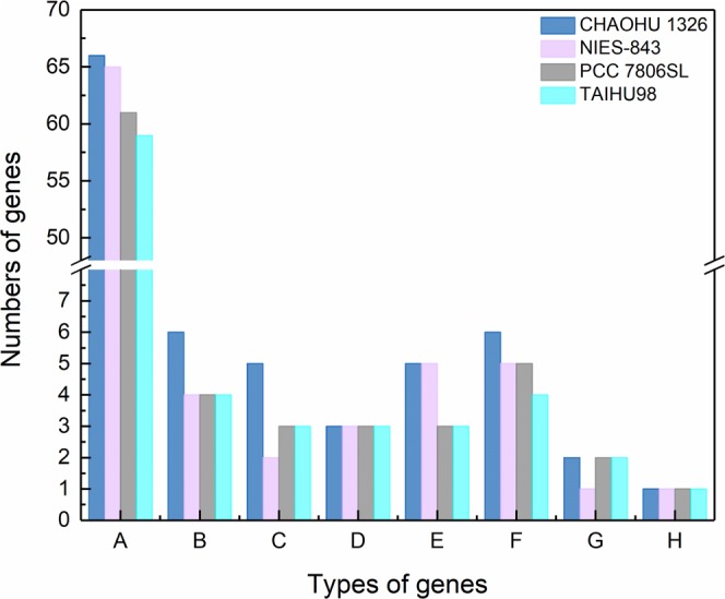
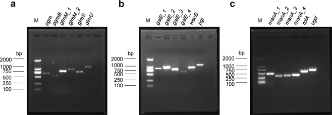
Microcystis, the dominant species among cyanobacterial blooms, normally forms colonies under natural conditions but exists as single cells or paired cells in axenic laboratory cultures after long-term cultivation. Here, a bloom-forming Microcystis aeruginosa strain CHAOHU 1326 was studied because it presents a colonial morphology and grows on the water surface during axenic laboratory culturing. We first examined the morphological features of strain CHAOHU 1326 and three other unicellular M. aeruginosa strains FACHB-925, FACHB-940, and FACHB-975 cultured under the same conditions by scanning and transmission electron microscopy. Then, we compared the extracellular polysaccharide (EPS)-producing ability of colonial strain CHAOHU 1326 to that of the three unicellular M. aeruginosa strains, and found that strain CHAOHU 1326 produced a higher amount of EPS than the other strains during growth. Moreover, based on genome sequencing, multiple gene clusters implicated in EPS biosynthesis and a cluster of 12 genes predicted to be involved in gas vesicle synthesis in strain CHAOHU 1326 were detected. These predicted genes were all functional and expressed in M. aeruginosa CHAOHU 1326 as determined by reverse transcription PCR. These findings provide a physiological and genetic basis to better understand colony formation and buoyancy control during M. aeruginosa blooming.


Conflict of interest statement

The authors declare no competing interests.

Figures

Figure 1
Figure 1
Neighbour-joining phylogenetic tree based on the 16S rRNA gene sequences of M. aeruginosa CHAOHU 1326 and other M. aeruginosa strains obtained from the GenBank database. Bootstrap values above 50% are shown at the branch nodes (1,000 replicates). Gloeothece membranacea PCC 6501 is used as outgroup. The scale bar represents 0.005 nucleotide substitutions per site. M. aeruginosa CHAOHU 1326 is indicated by the solid diamond (♦). Strains FACHB-925, FACHB-940, and FACHB-975 are marked by the empty diamond (◊).
Figure 2
Figure 2
Cultures of M. aeruginosa strains grown to early exponential phase in BG11 medium. Panels a–d show the cells of M. aeruginosa strains CHAOHU 1326, FACHB-925, FACHB-940, and FACHB-975, respectively. The macroscopic appearance of the M. aeruginosa cultures is shown in panel e, and the strains are marked by the numbers corresponding to their strain names. The small hole between colonies of strain CHAOHU 1326 is marked with an arrow in panel a.
Figure 3
Figure 3
SEM images of the four M. aeruginosa strains grown to mid-exponential phase in BG11 medium, including M. aeruginosa CHAOHU 1326 (a), M. aeruginosa FACHB-925 (b), M. aeruginosa FACHB-940 (c), and M. aeruginosa FACHB-975 (d).
Figure 4
Figure 4
TEM images of the four M. aeruginosa strains at mid-exponential growth phase in BG11 medium, including strains CHAOHU 1326 (a), FACHB-925 (b), FACHB-940 (c), and FACHB-975 (d). Poly-beta-hydroxybutyrate (PHB), poly granules (PH), lipid droplets (L), thylakoids (T), and gas vesicles (GV) are indicated.
Figure 5
Figure 5
Soluble EPS produced by the M. aeruginosa strains during growth in BG11 medium. The EPS yields are displayed by the columns and the growth curves of different strains are displayed by lines in panel a. The ratios of EPS yield to the dry cell weight of the different strains are shown in panel b. The molecular weight distribution of soluble EPS produced by M. aeruginosa CHAOHU 1326 is shown in panel c. Error bars represent standard deviation in at least triplicate. A statistical difference of p < 0.01 is marked with asterisks (**).
Figure 6
Figure 6
Types and numbers of genes encoding enzymes and proteins involved in extracellular polysaccharide biosynthesis and export in M. aeruginosa strains CHAOHU 1326, NIES-843, PCC 7806SL and TAIHU98. A: glycosyltransferases; B: isomerases; C: epimerases; D: phosphoglucomutases; E: glucose dehydrogenases; F: polysaccharide biosynthesis proteins; G: polysaccharide export proteins; H: bifunctional protein GlmU.
Figure 7
Figure 7
Transcriptional activity of genes involved in polysaccharide synthesis in M. aeruginosa CHAOHU 1326. The reverse transcription PCR products for pgm encoding an alpha-D-glucose phosphate-specific phosphoglucomutase, pgmB encoding a beta-phosphoglucomutase, glmM_1 encoding a phosphoglucosamine mutase, glmM_2 encoding a phosphoglucosamine mutase, glmS encoding a glutamine-fructose-6-phosphate aminotransferase, and glmU encoding bifunctional protein GlmU, are shown in panel a. Amplicons for galE_1 encoding a UDP-glucose 4-epimerase, galE_2 encoding a UDP-glucose 4-epimerase, galE_3 encoding a UDP-glucose 4-epimerase, galE_4 encoding a UDP-glucose 4-epimerase, wecB encoding a UDP-N-acetylglucosamine 2-epimerase, and pgi encoding a glucose-6-phosphate isomerase, are shown in panel b. Amplicons for manA_1 encoding a mannose-6-phosphate isomerase, manA_2 encoding a mannose-6-phosphate isomerase, manA_3 encoding a mannose-6-phosphate isomerase, manA_4 encoding a mannose-6-phosphate isomerase, rpiA encoding a ribose-5-phosphate isomerase, and ugd encoding a UDP-glucose 6-dehydrogenase are shown in panel c. (M) is a corresponding DNA molecular mass standard.
Figure 8
Figure 8
Organization and transcriptional analysis of genes involved in gas vesicle synthesis of M. aeruginosa CHAOHU 1326. The organization of the gvp gene cluster in M. aeruginosa CHAOHU 1326 is shown in panel a. The RT-PCR products of gvpA, gvpF/L, gvpN, and gvpW are shown in panel b, and the amplicons of gvpC, gvpG, gvpJ-X, gvpK, and gvpV are shown in panel c. (M) is a corresponding DNA molecular mass standard.

References

    1. Šejnohová, L. & Maršálek, B. In Ecology of Cyanobacteria II: Their Diversity in Space and Time (ed. Brian, A. W.) 195–228 (Springer Netherlands, 2012).
    1. Ndlela LL, Oberholster PJ, Wyk JHV, Cheng PH. An overview of cyanobacterial bloom occurrences and research in Africa over the last decade. Harmful Algae. 2016;60:11–26. doi: 10.1016/j.hal.201610001. - DOI - PubMed
    1. Paerl HW, Huisman J. Blooms like it hot. Science. 2008;320:57–58. doi: 10.1126/science.1155398. - DOI - PubMed
    1. Harke MJ, et al. A review of the global ecology, genomics, and biogeography of the toxic cyanobacterium, Microcystis spp. Harmful Algae. 2016;54:4–20. doi: 10.1016/j.hal.201512007. - DOI - PubMed
    1. Codd GA, Lindsay J, Young FM, Morrison LF, Metcalf JS. Harmful cyanobacteria. Aquatic Ecology. 2005;3:1–23. doi: 10.1007/1-4020-3022-3_1. - DOI

Publication types

Substances

Cite

AltStyle によって変換されたページ (->オリジナル) /