This site needs JavaScript to work properly. Please enable it to take advantage of the complete set of features!
Skip to main page content
U.S. flag

An official website of the United States government

Dot gov

The .gov means it’s official.
Federal government websites often end in .gov or .mil. Before sharing sensitive information, make sure you’re on a federal government site.

Https

The site is secure.
The https:// ensures that you are connecting to the official website and that any information you provide is encrypted and transmitted securely.

NIH NLM Logo
Log in
Access keys NCBI Homepage MyNCBI Homepage Main Content Main Navigation
. 2018 Jun 20;8(1):9229.
doi: 10.1038/s41598-018-27271-x.

Expression of human mutant cyclin dependent kinase 4, Cyclin D and telomerase extends the life span but does not immortalize fibroblasts derived from loggerhead sea turtle (Caretta caretta)

Affiliations

Expression of human mutant cyclin dependent kinase 4, Cyclin D and telomerase extends the life span but does not immortalize fibroblasts derived from loggerhead sea turtle (Caretta caretta)

Tomokazu Fukuda et al. Sci Rep. .

Abstract

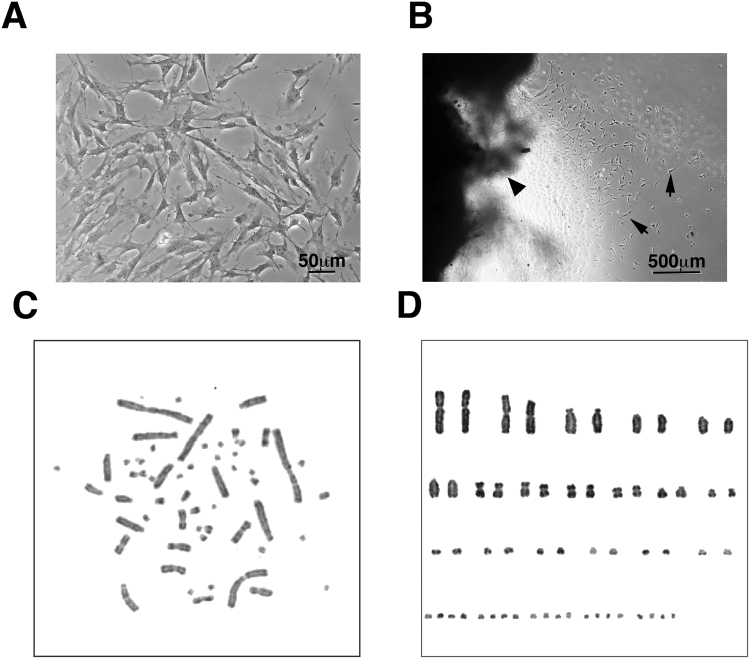
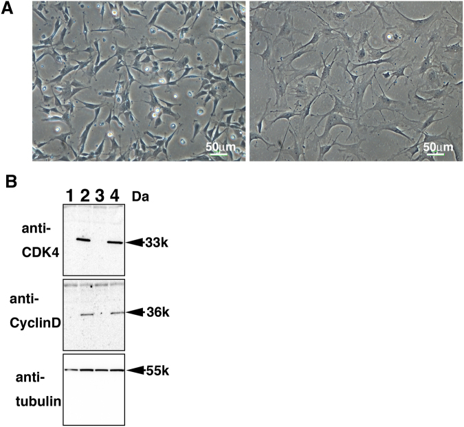
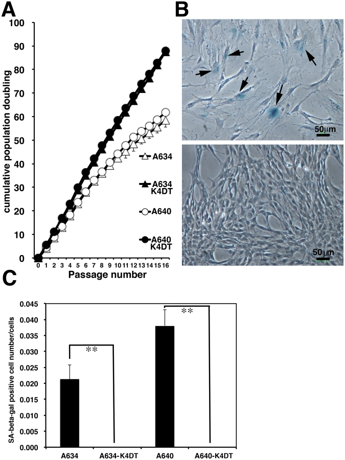
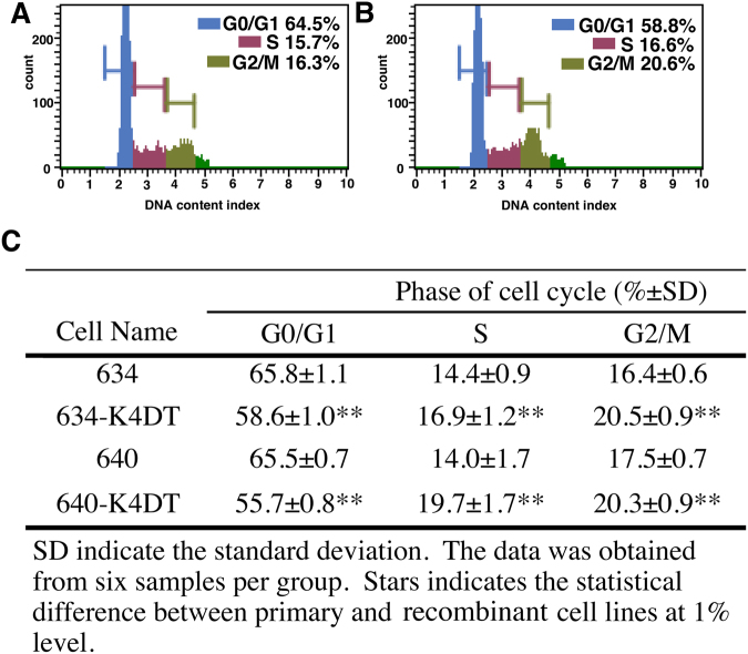
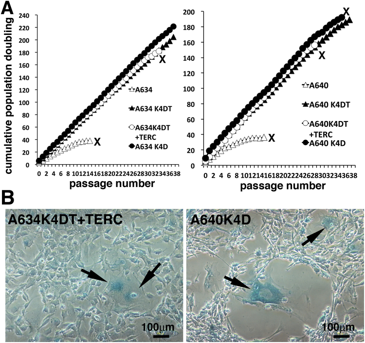
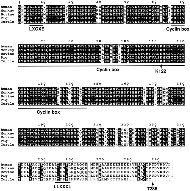
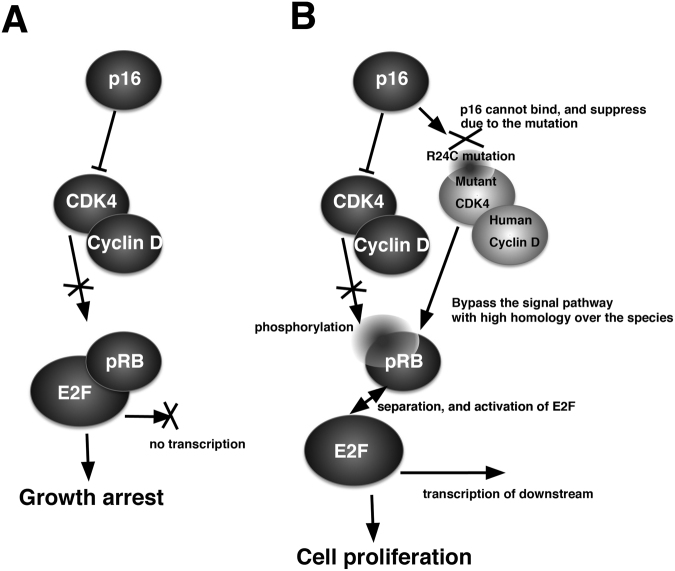
Conservation of the genetic resources of endangered animals is crucial for future generations. The loggerhead sea turtle (Caretta caretta) is a critically endangered species, because of human hunting, hybridisation with other sea turtle species, and infectious diseases. In the present study, we established primary fibroblast cell lines from the loggerhead sea turtle, and showed its species specific chromosome number is 2n = 56, which is identical to that of the hawksbill and olive ridley sea turtles. We first showed that intensive hybridization among multiple sea turtle species caused due to the identical chromosome number, which allows existence of stable hybridization among the multiple sea turtle species. Expressions of human-derived mutant Cyclin-dependent kinase 4 (CDK4) and Cyclin D dramatically extended the cell culture period, when it was compared with the cell culture period of wild type cells. The recombinant fibroblast cell lines maintained the normal chromosome condition and morphology, indicating that, at the G1/S phase, the machinery to control the cellular proliferation is evolutionally conserved among various vertebrates. To our knowledge, this study is the first to demonstrate the functional conservation to overcome the negative feedback system to limit the turn over of the cell cycle between mammalian and reptiles. Our cell culture method will enable the sharing of cells from critically endangered animals as research materials.


Conflict of interest statement

The authors declare no competing interests.

Figures

Figure 1
Figure 1
Primary cell culture of the loggerhead sea turtle under different conditions, and from frozen preserved tissue. (A) Cell morphology derived from the loggerhead sea turtle. Bar indicates 50 μm. (B) Primary cells obtained from the frozen preserved loggerhead tissue. Arrows indicate the primary cells. The arrowhead indicates the primary tissue. Bar indicates 500 μm. (C) Representative mitotic phase of the loggerhead cells. (D) Aligned chromosomes of the loggerhead cells. The result revealed a chromosome number of 2n = 56.
Figure 2
Figure 2
Introduction of enhanced green fluorescence protein (EGFP)-expressing retrovirus into primary cells of the loggerhead sea turtle. Left panels display EGFP detection of retrovirus-infected cells. Middle panels display cell morphology of retrovirus-infected cells, viewed under a differential interference contrast (DIC) microscope. Right panels display merged images of DIC and fluorescence images. Results with infection at 37 °C (A), 30 °C (B) and 26 °C (C).
Figure 3
Figure 3
Establishment of Cyclin-dependent kinase 4 (CDK4)-, Cyclin D- and telomerase-expressing cells derived from the loggerhead sea turtle. (A) left panel, cell morphology of the loggerhead-derived cells, expressing CDK4, Cyclin D, and telomerase. Right panel, cell morphology of the loggerhead primary cells. (B) Protein expression of the introduced CDK4 and Cyclin D. The expression of human CDK4, human Cyclin D, and endogenous tubulin were detected by using western blot analysis. Lane 1, primary A634 cell line; Lane 2, A634-K4DT cell line; Lane 3, primary A640 cell line; Lane 4, A640-K4DT cell line.
Figure 4
Figure 4
Growth characteristics and cellular senescence of primary and recombinant cells derived from the loggerhead sea turtle. (A) Sequential passage and cell growth of primary A634 cells, recombinant A634 derived K4DT cells, primary A640 cells and recombinant A640 derived K4DT cells. At the start of each passage, 5 ×ばつ 104 cells of each cell line were added to triplicate cell culture wells. (B) Detection of senescence-associated β-galactosidase staining in primary A634 cells (upper panel) and A634 derived K4DT cells (lower panel) after 16 passages. The arrows indicate positive blue staining, denoting cellular senescence. (C) Incidence of positively stained primary A634 cells, recombinant A634 derived K4DT cells, primary A640 cells and recombinant A640 derived K4DT cells. The percentage of positively stained cells counted in six randomly selected microscopic fields is shown in the graph. The double asterisks indicate statistical significance at the 1% level.
Figure 5
Figure 5
Cell cycle analysis of primary and recombinant cells derived from the loggerhead sea turtle. A and B, Histograms obtained from the cell cycle analysis: (A) primary cells (A634); and (B) recombinant cells (A634 derived K4DT). The percentages of cells in the G0/G1, S and G2/M phases are shown in color. (C) Percentages of primary and recombinant loggerhead-derived cells at each cell cycle phase. The data were obtained from five replicate samples. The double asterisks indicate statistical significance at the 1% level.
Figure 6
Figure 6
Detection of telomerase activity using stretch PCR. (A) Results of primary and recombinant sea turtle derived cell, Lane 1, Molecular weight marker, Lane 2, negative control, Lane 3, positive control sample (HeLa cells), Lane 4, A634 primary cells, Lane 5, A634 derived K4DT cells, Lane 6, A634 derived K4DT + TERC, Lane 7, A634 derived K4D cells. (B) Detection of telomerase activity with protein lysate (Lane 1), heat inactivated A634 derived protein (Lane 2), RNase treated A634 derived protein (Lane 3).
Figure 7
Figure 7
Sequential passage experiments with primary and recombinant cells. (A) Left panel, Results of sequential passages of A634 derived primary, A634 derived K4DT, A634 derived K4DT + TERC and A634 derived K4D cells. Right panel, Results of sequential passages of A640 derived primary, A640 derived K4DT, A640 derived K4DT + TERC and A640 derived K4D cells. X indicates the termination of cell growth. (B) Left panel, senescence-associated β-galactosidase staining of A634 derived K4DT + TERC cells at passage 36. Right panel, senescence-associated β-galactosidase staining of A640 derived K4D cells at passage 38. Arrows indicates the positively stained cells.
Figure 8
Figure 8
Multiple alignments of CDK4 protein among humans, monkeys, bovines, pigs, and turtles. The amino acid sequences were obtained from the UCSC Genome Bioinformatics database (http://www.ucsc.edu). The conserved amino acids are highlighted with black boxes. The functional motifs corresponding to the ATP-binding domain (A) and substrate-binding domain (S) are marked on the sequences. The core amino acid for the binding to p16 protein (24 R) is highlighted with an asterisk.
Figure 9
Figure 9
Multiple alignments of Cyclin D protein among humans, monkeys, bovines, pigs and turtles. The amino acid sequences were obtained from the UCSC Genome Bioinformatics database (http://www.ucsc.edu). The conserved amino acids are highlighted with black boxes. The functional motif of LXCXE (which is important for the binding with pRB), the Cyclin kinase box, and the LLXXXL motif (which is important for the binding with steroid receptor co-activators) are underlined.
Figure 10
Figure 10
Expected accelerated cell growth mechanism of mutant human-derived CDK4, Cyclin D, and TERT over the multiple species. (A) Cell growth arrest under the cellular stress. The protein level of p16 increases under the senescence. The p16 protein will bind to the pocket of the CDK4, and negatively regulates the activity of CDK4-Cyclin D complex. The inactivated CDK4-Cyclin D complex cannot induce the phosphorylation of pRB and its inactivation. Under the intact condition of pRB, E2F is not released from the binding status, resulting in no transcription of the downstream and growth arrest of the cells. (B) Enhanced cellular proliferation with mutant CDK4, Cyclin D and TERT. Due to the R24C mutation of mutant human-derived CDK4, p16 protein cannot suppress the activity of protein complex of mutant CDK4 and human Cyclin D. The exogenously introduced human-derived CDK4-Cyclin D complex has high homology with the endogenous CDK4-Cyclin D, and can form the protein complex with endogenous pRB, and phosphorylation. Due to the phosphorylation and inactivation of pRB, the transcription factor, E2F, would be released from the complex and induce cell proliferation.

References

    1. Barron MG. Ecological impacts of the deepwater horizon oil spill: implications for immunotoxicity. Toxicologic pathology. 2012;40:315–320. doi: 10.1177/0192623311428474. - DOI - PubMed
    1. Lu Y, et al. Establishment and characterization of 13 cell lines from a green turtle (Chelonia mydas) with fibropapillomas. In Vitro Cell Dev Biol Anim. 1999;35:389–393. doi: 10.1007/s11626-999-0113-6. - DOI - PubMed
    1. Herbst LH, Moretti JE, Brown R, Sundberg T, Klein JP. PA. Experimental transmission of green turtle fibropapillomatosis using cell-free tumor extracts. Diseases of Aquatic Organisms. 1995;22:1–12. doi: 10.3354/dao022001. - DOI
    1. Lara-Ruiz P, Lopez GG, Santos FR, Soares LS. Extensive hybridization in hawksbill turtles (Eretmochelys imbricata) nesting in Brazil revealed by mtDNA analyses. Conservation Genetics. 2006;7:773–781. doi: 10.1007/s10592-005-9102-9. - DOI
    1. Kamezaki N. The possibility of hybridization between the loggerhead turtle, Caretta caretta, and the hawksbill turtle, Eretmochelys imbricata, in specimens hatched from eggs collected in Chita Peninsula. Japanese Journal of Herpetology. 1983;10:52–53.

Publication types

MeSH terms

Cite

AltStyle によって変換されたページ (->オリジナル) /