This site needs JavaScript to work properly. Please enable it to take advantage of the complete set of features!
Skip to main page content
U.S. flag

An official website of the United States government

Dot gov

The .gov means it’s official.
Federal government websites often end in .gov or .mil. Before sharing sensitive information, make sure you’re on a federal government site.

Https

The site is secure.
The https:// ensures that you are connecting to the official website and that any information you provide is encrypted and transmitted securely.

NIH NLM Logo
Log in
Access keys NCBI Homepage MyNCBI Homepage Main Content Main Navigation
. 2016 Oct;113(10):2088-99.
doi: 10.1002/bit.25976. Epub 2016 Mar 28.

A new paradigm for producing astaxanthin from the unicellular green alga Haematococcus pluvialis

Affiliations

A new paradigm for producing astaxanthin from the unicellular green alga Haematococcus pluvialis

Zhen Zhang et al. Biotechnol Bioeng. 2016 Oct.

Abstract

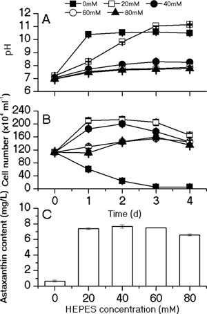
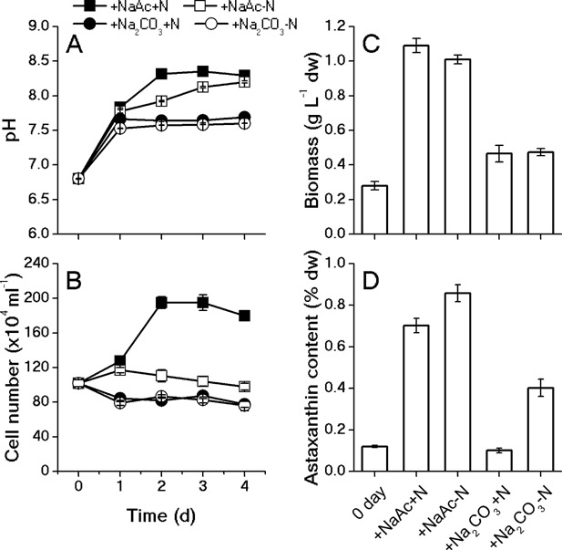
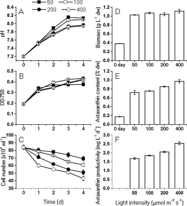
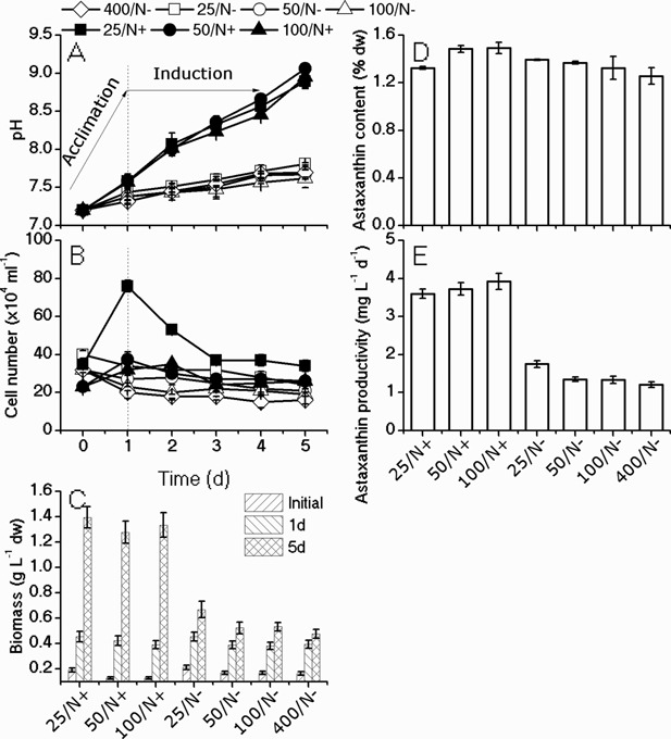
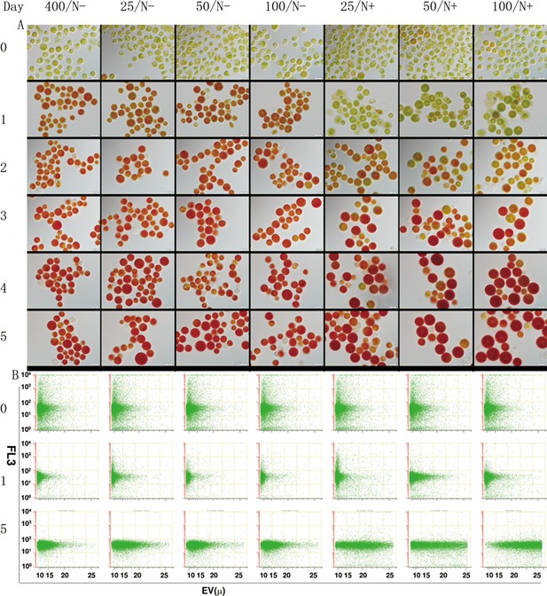
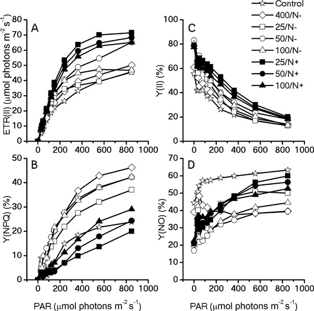
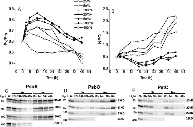
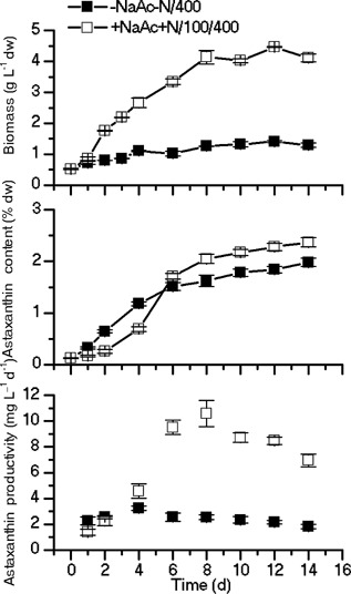
The unicellular green alga Haematococcus pluvialis has been exploited as a cell factory to produce the high-value antioxidant astaxanthin for over two decades, due to its superior ability to synthesize astaxanthin under adverse culture conditions. However, slow vegetative growth under favorable culture conditions and cell deterioration or death under stress conditions (e.g., high light, nitrogen starvation) has limited the astaxanthin production. In this study, a new paradigm that integrated heterotrophic cultivation, acclimation of heterotrophically grown cells to specific light/nutrient regimes, followed by induction of astaxanthin accumulation under photoautotrophic conditions was developed. First, the environmental conditions such as pH, carbon source, nitrogen regime, and light intensity, were optimized to induce astaxanthin accumulation in the dark-grown cells. Although moderate astaxanthin content (e.g., 1% of dry weight) and astaxanthin productivity (2.5 mg L(-1) day(-1) ) were obtained under the optimized conditions, a considerable number of cells died off when subjected to stress for astaxanthin induction. To minimize the susceptibility of dark-grown cells to light stress, the algal cells were acclimated, prior to light induction of astaxanthin biosynthesis, under moderate illumination in the presence of nitrogen. Introduction of this strategy significantly reduced the cell mortality rate under high-light and resulted in increased cellular astaxanthin content and astaxanthin productivity. The productivity of astaxanthin was further improved to 10.5 mg L(-1) day(-1) by implementation of such a strategy in a bubbling column photobioreactor. Biochemical and physiological analyses suggested that rebuilding of photosynthetic apparatus including D1 protein and PsbO, and recovery of PSII activities, are essential for acclimation of dark-grown cells under photo-induction conditions. Biotechnol. Bioeng. 2016;113: 2088-2099. © 2016 The Authors. Biotechnology and Bioengineering Published by Wiley Periodicals, Inc.

Keywords: Haematococcus pluvialis; acclimation; astaxanthin; heterotrophy.


Figures

Figure 1
Figure 1
Effects of different concentrations of HEPES buffer on pH (A), cell number (B), and astaxanthin content (C). The pH and cell numbers were monitored on a daily basis during 4‐day induction; astaxanthin content were measured on day 4. Values represent the mean ± S.D (n = 3).
Figure 2
Figure 2
Effects of carbon source and nitrate on the pH (A), cell number (B) and biomass (C), astaxanthin content (D). The pH and cell number were monitored on a daily basis during 4‐day induction; biomass and astaxanthin content were measured on day 4.
Figure 3
Figure 3
Effects of light intensities (50, 100, 200, 400 μmol photons m−2 s−1) on the pH (A), OD750 (B), cell number (C) and biomass (D), astaxanthin content (E) and astaxanthin productivity (F). The pH, OD, and cell numbers were monitored on a daily basis during 4‐day induction; biomass, astaxanthin content and productivity were measured on day 4.
Figure 4
Figure 4
Effects of acclimation under moderate light intensity with/without nitrate on the pH (A), cell number changes (B) in the 4 days induction, and biomass (C), astaxanthin content (D) and astaxanthin productivity (E) at the end of the 5 days procession. Dark‐grown algal cells were acclimated for 1 day under moderate light (25, 50, 100 μmol photons m−2 s−1) and N‐replete (/N+) or N‐depleted (/N−) conditions, before being subjected to high‐light (400 μmol photons m−2 s−1) and N‐depleted induction for 4 days. The acclimated cells were compared to those subjected to induction without acclimation (400/N−).
Figure 5
Figure 5
Microscopic observation (upper panel, A) and flow cytometry analysis (bottom panel, B) of cellular morphological changes during the acclimation and induction processes. Dark‐grown cells (day 0) were acclimated under moderate light (25, 50, 100 μmol photons m−2 s−1) and N‐replete (/N+) or N‐depleted (/N−) conditions for 1 day, and then subjected to HL and N‐depleted for 4 days (days 2–5) to induce astaxanthin accumulation. The cell size (EV, electronic volume) and chlorophyll fluorescence (FL3) measured from 10,000 cells by using a flow cytometer were displayed in the plots.
Figure 6
Figure 6
Light response curves of the chlorophyll fluorometry parameters for the acclimated algae cells. Chlorophyll fluorescence were obtained with a series of photosynthetically active radiances (0, 30, 37, 46, 77, 119, 150, 240, 363, 555, 849 μmol photons m−2 s−1) to calculate (A) electron transport rate, (B) Y(NPQ), (C) Y(II), and (D) Y(NO).
Figure 7
Figure 7
Changes in chlorophyll fluorometry parameters during the acclimation and induction processes (A and B) and western blot of protein PsbA, PsbO, and PetC (C, D, and E, respectively). The actinic lights for chlorophyll fluorometry analysis were set at 400 μmol photons m−2 s−1. The algae cells were collected and adapted in dark for 30 min before measurements. The samples for western blot were taken every 12 h during the 2‐day acclimation and induction.
Figure 8
Figure 8
Biomass and astaxanthin content of H. pluvialis cultured in the 1 L bubbling column photobioreactor. —▪— dark‐grown cells were transferred to the N‐depleted growth medium and subjected to the illumination of 400 μmol photons m−2 s−1 for the astaxanthin induction, —しろいしかく— cells were acclimated under N replete (2 mM) and 100 μmol photons m−2 s−1 conditions prior to the induction conditions under N deplete and 400 μmol photons m−2 s−1.

References

    1. Aflalo C, Meshulam Y, Zarka A, Boussiba S. 2007. On the relative efficiency of two‐vs. one‐stage production of astaxanthin by the green alga Haematococcus pluvialis . Biotechnol Bioeng 98(1):300–305. - PubMed
    1. Armstrong GA, Runge S, Frick G, Sperling U, Apel K. 1995. Identification of NADPH: protochlorophyllide oxidoreductases A and B: A branched pathway for light‐dependent chlorophyll biosynthesis in Arabidopsis thaliana . Plant Physiol 108(4):1505–1517. - PMC - PubMed
    1. Baker NR. 2008. Chlorophyll fluorescence: A probe of photosynthesis in vivo. Annu Rev Plant Biol 59:89–113. - PubMed
    1. Björkman O, Demmig B. 1987. Photon yield of O2 evolution and chlorophyll fluorescence characteristics at 77K among vascular plants of diverse origins. Planta 170(4):489–504. - PubMed
    1. Boussiba S, Vonshak A. 1991. Astaxanthin accumulation in the green alga Haematococcus pluvialis . Plant Cell Physiol 32(7):1077–1082.

Publication types

MeSH terms

Cite

AltStyle によって変換されたページ (->オリジナル) /