This site needs JavaScript to work properly. Please enable it to take advantage of the complete set of features!
Skip to main page content
U.S. flag

An official website of the United States government

Dot gov

The .gov means it’s official.
Federal government websites often end in .gov or .mil. Before sharing sensitive information, make sure you’re on a federal government site.

Https

The site is secure.
The https:// ensures that you are connecting to the official website and that any information you provide is encrypted and transmitted securely.

NIH NLM Logo
Log in
Access keys NCBI Homepage MyNCBI Homepage Main Content Main Navigation
. 2016 Jul 13:7:1040.
doi: 10.3389/fpls.2016.01040. eCollection 2016.

Concanavalin A Disrupts the Release of Fibrous Material Necessary for Zygote Formation of a Unicellular Charophycean Alga, Closterium peracerosum-strigosum-littorale Complex

Affiliations

Concanavalin A Disrupts the Release of Fibrous Material Necessary for Zygote Formation of a Unicellular Charophycean Alga, Closterium peracerosum-strigosum-littorale Complex

Jun Abe et al. Front Plant Sci. .

Abstract

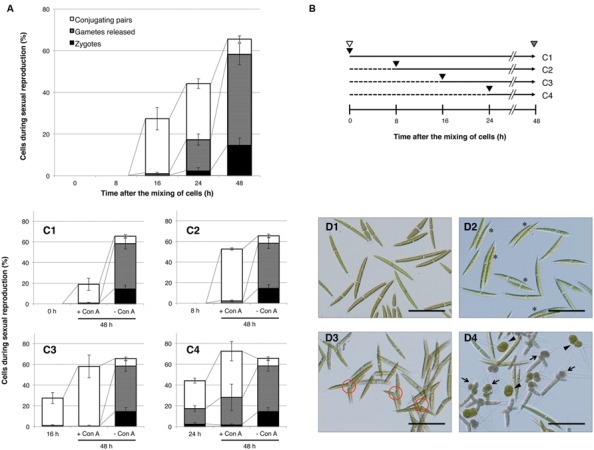
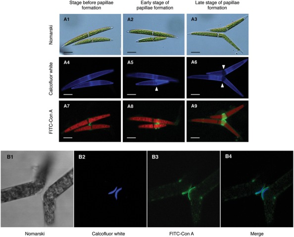
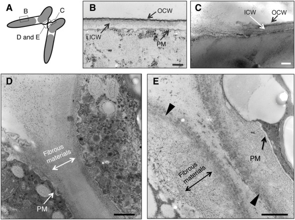
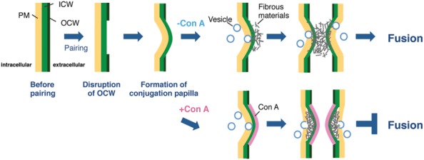
The Closterium peracerosum-strigosum-littorale (C. psl.) complex is the best characterized charophycean alga with respect to the processes of sexual reproduction. We examined the effect of concanavalin A (Con A) on physiological and ultrastructural changes during the conjugation of the C. psl. complex. Two heterothallic gametangial cells formed a sexual pair as usual; however, the release of gametes was completely blocked by the addition of Con A. Fluorescein isothiocyanate-labeled Con A bound to the outermost layer of the conjugation papillae of paired cells. In the absence of Con A, the disruption of outer cell walls on the conjugation papillae and the secretion of fibrous materials from the conjugation papillae were observed using a transmission electron microscope, but Con A-treated cells did not show these changes. Instead, a highly electron-dense layer was observed in the outermost papillae, and the excess fibrous materials remained at the inside of the layer. These results suggest that an unknown molecule(s) recognized by Con A is essential for the diffusion of fibrous materials at the conjugation papillae, which is an indispensable step for gamete release during conjugation of the C. psl. complex.

Keywords: Closterium; Concanavalin A (Con A); charophycean; conjugation; gamete; sexual reproduction.


Figures

FIGURE 1
FIGURE 1
Progress of sexual reproduction in the presence or absence of concanavalin A (Con A). (A) Sexual reproductive stages in the absence of Con A. Independently prepared sexually differentiated mt+ and mt- cells were mixed and incubated. At intervals shown on the x-axis, cells undergoing sexual reproduction (conjugating pair-forming cells, gamete-releasing cells, and zygotes) were counted (n = 3). (B) The experimental procedure to determine the developmental stages that are inhibited by Con A. Independently prepared sexually differentiated mt+ and mt- cells were mixed (shown by white arrowheads) and incubated for 48 h. At 0, 8, 16, or 24 h after the mixing, Con A was added to the respective test tubes (black arrowhead). Populations of sexually reproductive cells were examined at 48 h after the mixing (gray arrowhead). Horizontal arrows with solid and dashed lines indicate periods of incubation with or without Con A, respectively. The numbers on the right correspond to the results shown in (C1–4). (C1–4) Effect of Con A on the progress of sexual reproduction. Con A was added at 0 (C1), 8 (C2), 16 (C3), or 24 h (C4) after the mixing of cells. The bar on the left in each graph shows the cell populations at a time of Con A addition. Bars at the center and right in each graph indicate the cell population after 48 h of incubation with or without Con A, respectively. White, gray, and black boxes show the cell populations in conjugating pairs, gamete-releasing cells, and zygotes, respectively. (D1–4) Photographs of mixed cells with or without Con A. D1: Initial cells (0 h after the mixing of cells). D2: Conjugating pairs (16 h after the mixing of cells). Conjugating pairs are shown by asterisks. D3: Cells after 48 h of incubation in the presence of Con A (Con A was added 8 h after mixing of the cells). Conjugation papillae are shown in red circles. D4: Cells after 48 h of incubation in the absence of Con A. Gamete-releasing cells and zygotes are shown by arrows and arrowheads, respectively. Scale bar: 100 μm.
FIGURE 2
FIGURE 2
Subcellular localization of Con A-binding molecule(s). (A1–9) Development of conjugation papillae and subcellular localization of Con A binding molecule(s) during development. Both mating type cells were separately pulse-labeled by Calcofluor White prior to the mixing of cells. Fluorescein isothiocyanate (FITC)-labeled Con A was added to the mixed cells just before pair formation (8 h after the mixing of cells). (A1–3) Nomarski images of three developmental stages of papilla formation. (A4–6) Calcofluor White staining images. Novel unstained papillae (arrowhead) expanded during development. (A7–9) Localization of FITC-Con A binding molecule(s). Red signals in each cell represent auto-fluorescence of chlorophyll. Scale bar: 20 μm. (B1–4) Localization of Con A binding molecule(s) on conjugation papillae. Calcofluor White and FITC-labeled Con A were simultaneously added to mixed cells extruding the conjugation papillae. (B1) Nomarski image of conjugation papillae. (B2) Fluorescence by Calcofluor White. (B3) Fluorescence by FITC-labeled Con A. (B4) Merged image of B2 and B3.
FIGURE 3
FIGURE 3
Ultrastructural observations of paired cells using a transmission electron microscope (TEM). (A) Illustration of TEM sections observed in this study. (B) Closterium cell wall consists of two layers: outer cell wall (OCW) and inner cell wall (ICW). Around the conjugation papillae, the OCW was disrupted (C) and fibrous-like materials were released from the papillae (D). When Con A was added prior to cell fusion (16 h after the mixing), the fibrous-like materials accumulated in the conjugation papillae of respective paired cells. Increased electron density at the outermost layer was also observed (arrowhead) (E). PM, plasma membrane. Bars: 250 nm (B), 200 nm (C), and 500 nm (D,E).
FIGURE 4
FIGURE 4
Possible model for cell fusion in the C. psl. Complex. Once mt+ and mt- cells have formed a conjugation pair, the OCWs (OCW) at the point of adhesion of the two cells are disrupted. The cytoplasm of both paired cells is extruded from the thin wall area to form papillae by osmotic pressure. In parallel, fibrous materials, which are indispensable for gamete release, diffuse from the ICW to the tip of papillae with the supply of materials by vesicle transport. The target molecule of Con A is synthesized at adhesion sites prior to the formation of papillae and finally acts to unfold or refold the fibrous materials on the surface of papillae. In the presence of Con A, the highly electron-dense layer at the outermost papillae is appeared and the unfolding or refolding by the target molecule is inhibited; therefore, paired cells cannot complete gamete release and cell fusion.

References

    1. Abe J., Hori S., Tsuchikane Y., Kitao N., Kato M., Sekimoto H. (2011). Stable nuclear transformation of the Closterium peracerosum-strigosum-littorale complex. Plant Cell Physiol. 52 1676–1685. 10.1093/pcp/pcr103 - DOI - PubMed
    1. Akatsuka S., Sekimoto H., Iwai H., Fukumoto R., Fujii T. (2003). Mucilage secretion regulated by sex pheromones in Closterium peracerosum-strigosum-littorale complex. Plant Cell Physiol. 44 1081–1087. 10.1093/pcp/pcg131 - DOI - PubMed
    1. Akatsuka S., Tsuchikane Y., Fukumoto R., Fujii T., Sekimoto H. (2006). Physiological characterization of the sex pheromone protoplast-release-inducing protein from the Closterium peracerosum-strigosum-littorale complex (Charophyta). Phycol. Res. 54 116–121. 10.1111/j.1440-1835.2006.00418.x - DOI
    1. Hirano N., Marukawa Y., Abe J., Hashiba S., Ichikawa M., Tanabe Y., et al. (2015). A Receptor-like kinase, related with cell wall sensor of higher plants, is required for sexual reproduction in the unicellular charophycean alga, Closterium peracerosum-strigosum-littorale complex. Plant Cell Physiol. 56 1456–1462. 10.1093/pcp/pcv065 - DOI - PubMed
    1. Hori S., Sekimoto H., Abe J. (2012). Properties of cell surface carbohydrates in sexual reproduction of the Closterium peracerosum–strigosum–littorale complex (Zygnematophyceae, Charophyta). Phycol. Res. 60 254–260. 10.1111/j.1440-1835.2012.00656.x - DOI

LinkOut - more resources

Cite

AltStyle によって変換されたページ (->オリジナル) /