This site needs JavaScript to work properly. Please enable it to take advantage of the complete set of features!
Skip to main page content
U.S. flag

An official website of the United States government

Dot gov

The .gov means it’s official.
Federal government websites often end in .gov or .mil. Before sharing sensitive information, make sure you’re on a federal government site.

Https

The site is secure.
The https:// ensures that you are connecting to the official website and that any information you provide is encrypted and transmitted securely.

NIH NLM Logo
Log in
Access keys NCBI Homepage MyNCBI Homepage Main Content Main Navigation
. 2015 Dec 1;10(12):e0143473.
doi: 10.1371/journal.pone.0143473. eCollection 2015.

Immortalization of Fetal Bovine Colon Epithelial Cells by Expression of Human Cyclin D1, Mutant Cyclin Dependent Kinase 4, and Telomerase Reverse Transcriptase: An In Vitro Model for Bacterial Infection

Affiliations

Immortalization of Fetal Bovine Colon Epithelial Cells by Expression of Human Cyclin D1, Mutant Cyclin Dependent Kinase 4, and Telomerase Reverse Transcriptase: An In Vitro Model for Bacterial Infection

Kengo Kuroda et al. PLoS One. .

Abstract

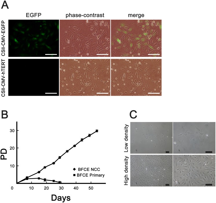
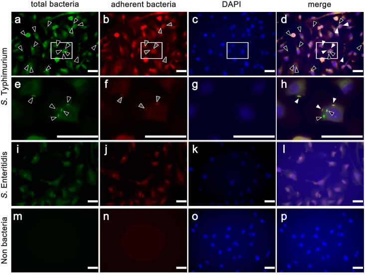
Cattle are the economically important animals in human society. They are essential for the production of livestock products such as milk and meats. The production efficiency of livestock products is negatively impacted by infection with zoonotic pathogens. To prevent and control infectious diseases, it is important to understand the interaction between cattle tissue and pathogenic bacteria. In this study, we established an in vitro infection model of an immortalized bovine colon-derived epithelial cell line by transducing the cells with lentiviral vectors containing genes encoding cell cycle regulators cyclin D1, mutant cyclin dependent kinase 4 (CDK4), and human telomerase reverse transcriptase (TERT). The established cell line showed continuous cell proliferation, expression of epithelial markers, and an intact karyotype, indicating that the cells maintained their original nature as colon-derived epithelium. Furthermore, we exposed the established cell line to two strains of Salmonella enterica and EHEC. Interestingly, S. Typhimurium showed higher affinity for the established cell line and invaded the cytoplasm than S. Enteritidis. Quantitative RT-PCR revealed that gene expression of Toll-like receptor 1 (TLR1), TLR 2 and TLR 3, whereas TLR 4, 5 and 6 were not detectable in established cells. Our established immortalized colon-derived epithelial cell should be a useful tool for studies evaluating the molecular mechanisms underlying bacterial infection.


Conflict of interest statement

Competing Interests: The authors have declared that no competing interests exist.

Figures

Fig 1
Fig 1. The efficiency of virus infection, cell morphology, and population doubling of the established BFCE-K4DT cells.
(A) Green fluorescence was detectable in primary cells transfected with CSII-CMV-EGFP vector (upper panels), while cells transfected with CSII-CMV-TERT vector were not fluoresce (lower panels). Scale bars: 50 μm. (B) The results of the population doubling assay in BFCE primary cells (squares) and BFCE-K4DT cells (diamonds) were plotted. Experiments were carried out in triplicate, and the averages and standard deviations (SD) were calculated. (C) The morphologies of the established cell line after cloning (after 15 times passage). BFCE-K4DT cells show an epithelial-like rounded shape in low-density culture (upper panels) and sheet-like morphology in high-density culture (lower panels). Scale bars: 100 μm.
Fig 2
Fig 2. Results of the immunoblotting analysis and TRAP assay.
(A) Detection of CDK4 and cyclin D1 by immunoblotting. Lane 1, BFCE primary cells; Lanes 2 and 3, BFCE-K4DT cells. (B) Detection of telomerase activity by TRAP assay. Lane 1, 1 kb ladder marker; lane 2, CHAPS buffer as a negative control; lane 3, 293T cells as a positive control; lane 4, BFCE primary cells; lane 5 and 6, BFCE-K4DT cells. The 6-bp ladders were detectable in lanes of positive control and BFCE-K4DT cells. Each experiment was performed in duplicates after cloning (primary cells: 2 times passage, K4DT cells: 15 times passage) and representative data were shown here.
Fig 3
Fig 3. Karyotype analysis of established BFCE-K4DT cells.
All mitotic chromosome spreads (50 of 50 examined) from BFCE-K4DT cells showed a 2n = 60XY pattern. This analysis was performed by using cloned cells after 15 times passage.
Fig 4
Fig 4. Fluorescent immunohistochemical staining for detection of epithelial markers.
BFCE-K4DT cells (passage number = 15) and BFCE-Primary cells (passage number = 2) were positive for E-cadherin and Cytokeratin-8, whereas BFF NCC cells (passage number was unknown) were negative for both markers. Scale bars: 50 μm. Each staining was carried for 3 times and representative pictures were shown here.
Fig 5
Fig 5. Fluorescence microscopic images of cells subjected to the adhesion and invasion assay.
S. Typhimurium (a-d: low-power fields and e-h; high-power fields), S. Enteritidis (i-l) after infection, and non-infected control (m-p)) in BFCE-K4DT cells (passage number = 15). Total bacteria were stained green (a, e, i, and m; black arrows), adherent bacteria were stained red (b, f, j, n; white arrows), nuclei of cells were stained blue, and merge images of extracellular bacteria (stars) and intracellular bacteria (black arrows) appear yellow and green respectively (d, h, l, p). Each staining was carried for 3 times and representative pictures were shown here.
Fig 6
Fig 6. Quantification of bacterial adhesion and invasion and SEM microscopy.
(A) Total S. Enteritidis or S. Typhimurium were harvested after each infection time (30, 60, 120 min) and plated in agar dishes to count the colony number (gray bars). Invasive bacteria were also harvested after 2 h gentamycin treatment at the each time point and count the colony by same procedures as total bacteria (black bars). (B) EHEC adhesion to BFCE-Primary cells (gray bars) and BFCE-K4DT cells (black bars) were also quantification by same methods as Salmonella infection. (C) SEM observations revealed the adhesion of S. Enteritidis (a), S. Typhimurium (b) and EHEC (c) to established BECE-K4DT cells (bars: 10 μm). Experiments were carried out in triplicate, and the averages and standard deviations (SD) were calculated (A and B) and representative figures were shown (C).

References

    1. Ferens WA, Hovde CJ. Escherichia coli O157:H7: animal reservoir and sources of human infection. Foodborne Pathog Dis. 2011;8(4):465–87. Epub 2010年12月02日. 10.1089/fpd.2010.0673 - DOI - PMC - PubMed
    1. Costa LF, Paixao TA, Tsolis RM, Baumler AJ, Santos RL. Salmonellosis in cattle: advantages of being an experimental model. Res Vet Sci. 2012;93(1):1–6. Epub 2012年04月10日. 10.1016/j.rvsc.201203002 . - DOI - PubMed
    1. Rosselin M, Abed N, Virlogeux-Payant I, Bottreau E, Sizaret PY, Velge P, et al. Heterogeneity of type III secretion system (T3SS)-1-independent entry mechanisms used by Salmonella Enteritidis to invade different cell types. Microbiology. 2011;157(Pt 3):839–47. Epub 2010年11月27日. 10.1099/mic.0.044941-0 . - DOI - PubMed
    1. Ozer HL, Banga SS, Dasgupta T, Houghton J, Hubbard K, Jha KK, et al. SV40-mediated immortalization of human fibroblasts. Exp Gerontol. 1996;31(1–2):303–10. Epub 1996年01月01日. . - PubMed
    1. Jha KK, Banga S, Palejwala V, Ozer HL. SV40-Mediated immortalization. Exp Cell Res. 1998;245(1):1–7. Epub 1998年11月26日. 10.1006/excr.1998.4272 . - DOI - PubMed

MeSH terms

LinkOut - more resources

Cite

AltStyle によって変換されたページ (->オリジナル) /