This site needs JavaScript to work properly. Please enable it to take advantage of the complete set of features!
Skip to main page content
U.S. flag

An official website of the United States government

Dot gov

The .gov means it’s official.
Federal government websites often end in .gov or .mil. Before sharing sensitive information, make sure you’re on a federal government site.

Https

The site is secure.
The https:// ensures that you are connecting to the official website and that any information you provide is encrypted and transmitted securely.

NIH NLM Logo
Log in
Access keys NCBI Homepage MyNCBI Homepage Main Content Main Navigation
Review
. 2022 Apr 30;23(9):5028.
doi: 10.3390/ijms23095028.

When the Locus Coeruleus Speaks Up in Sleep: Recent Insights, Emerging Perspectives

Affiliations
Review

When the Locus Coeruleus Speaks Up in Sleep: Recent Insights, Emerging Perspectives

Alejandro Osorio-Forero et al. Int J Mol Sci. .

Abstract

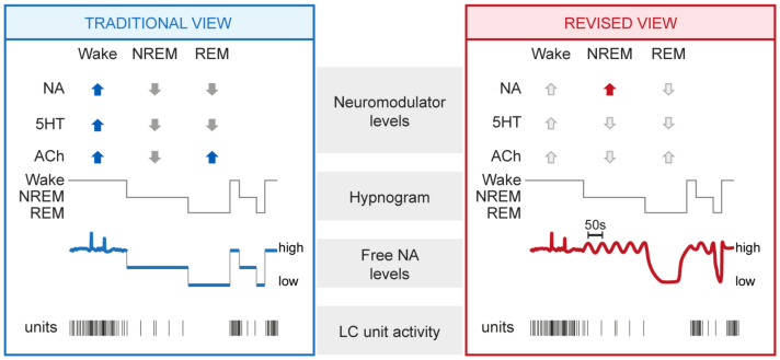
For decades, numerous seminal studies have built our understanding of the locus coeruleus (LC), the vertebrate brain's principal noradrenergic system. Containing a numerically small but broadly efferent cell population, the LC provides brain-wide noradrenergic modulation that optimizes network function in the context of attentive and flexible interaction with the sensory environment. This review turns attention to the LC's roles during sleep. We show that these roles go beyond down-scaled versions of the ones in wakefulness. Novel dynamic assessments of noradrenaline signaling and LC activity uncover a rich diversity of activity patterns that establish the LC as an integral portion of sleep regulation and function. The LC could be involved in beneficial functions for the sleeping brain, and even minute alterations in its functionality may prove quintessential in sleep disorders.

Keywords: Alzheimer’s disease; NREM sleep; REM sleep; arousability; infraslow time scale; microvasculature; monoamine; noradrenaline; sleep architecture; sleep disorder.


Conflict of interest statement

The authors declare no conflict of interest.

Figures

Figure 1
Figure 1
Summary of traditional and revised views on the neuromodulatory profiles of wakefulness and sleep, with a focus on noradrenergic signaling. Traditional (left) and revised (right) views derived from animal experimentation are summarized and complemented with data-derived schematic representations of NA dynamics and LC unit activity. From top to bottom: mean levels of major neuromodulators (blue up and gray down arrows symbolize high and low levels in the traditional view), a representative hypnogram of mouse sleep–wake behavior, free NA levels, and representative discharges of a LC unit. Novel insights central to the revised view are highlighted with the red arrow, whereas unaltered neuromodulatory levels are shown with light grey arrows. NA, noradrenaline; 5HT, serotonin; ACh, acetylcholine; NREM, NREM sleep; REM, REM sleep.
Figure 2
Figure 2
Real-time dynamics of NA levels in somatosensory thalamus, forebrain sleep spindle power, and heart rate during NREM sleep. Representative simultaneous recordings in a freely behaving mouse combining (from top to bottom): hypnogram (gray), free NA levels in somatosensory thalamus obtained through fiberphotometry imaging (red, A1), local field potential sigma power (10–15 Hz) in somatosensory cortex (dark blue, A2) and heart rate (light blue, A3), with labeled portions (A, B, C) shown expanded on the right. The variations in sigma power reflect the clustering of sleep spindle density ([72]). Insets on the right expand portions of the traces highlighted with letters in the hypnogram to show (A) NREM sleep (double-headed arrow marks the 50 s periodicity); (B) NREM-to-REM sleep transitions (double-headed arrow marks the decay time of NA levels prior to REM sleep onset); (C) NREM-to-wake transitions. Portions of two of these traces have been published previously [72]. NA, noradrenaline; W, wakefulness; NR, NREM sleep; R, REM sleep; ΔF/F, relative fluorescence changes; AU, arbitrary unit; bpm, beats per minute.
Figure 3
Figure 3
Perspectives for the implication of the LC in healthy and disrupted sleep. Schematic indicating the types of signals monitored by the LC and the implications of LC function and dysfunction for sleep. (A) The LC monitors external stimuli (e.g., sensory stimuli such as touch or sound, symbolized by a feather and a musical note, see Section 4.1), internal stimuli (symbolized by the heart, see Section 4.5), and internal brain states important for the regulation of NREM-to-REM sleep transitions (symbolized by the brain, see Section 3.4 and Section 4.3). (B) Depending on the LC status (healthy or damaged), beneficial or adverse consequences on sleep can arise. Several outcomes are listed on the right. The LC micrograph was obtained from an immunohistochemically stained brain section of one of the mice used for the data published in [72]. The color choice of cell labeling was made deliberately to mark it as the sky-blue spot. The blurring of the blue color in the bottom micrograph symbolizes both structural and functional alterations that can lead to LC dysfunction.

References

    1. Manger P.R., Eschenko O. The Mammalian Locus Coeruleus Complex—Consistencies and Variances in Nuclear Organization. Brain Sci. 2021;11:1486. doi: 10.3390/brainsci11111486. - DOI - PMC - PubMed
    1. Fuxe D. Evidence for existence of monoamine-containing neurons in central nervous system. I. Demonstration of monoamines in the cell bodies of brain stem neurons. Acta Physiol. Scand. 1964;62:1–55. - PubMed
    1. Maeda T., Pin C., Salvert D., Ligier M., Jouvet M. Les neurones contenant des catecholamines du tegmentum pontique et leurs voies de projection chez le chat. Brain Res. 1973;57:119–152. doi: 10.1016/0006-8993(73)90572-6. - DOI - PubMed
    1. Bogerts B. A brainstem atlas of catecholaminergic neurons in man, using melanin as a natural marker. J. Comp. Neurol. 1981;197:63–80. doi: 10.1002/cne.901970106. - DOI - PubMed
    1. Robertson S.D., Plummer N.W., De Marchena J., Jensen P. Developmental origins of central norepinephrine neuron diversity. Nat. Neurosci. 2013;16:1016–1023. doi: 10.1038/nn.3458. - DOI - PMC - PubMed
Cite

AltStyle によって変換されたページ (->オリジナル) /