Comment
doi: 10.1073/pnas.2204155119.
Epub 2022 Apr 22.
Time and tide of cerebellar synchrony
Affiliations
- PMID: 35452313
- PMCID: PMC9170046
- DOI: 10.1073/pnas.2204155119
Item in Clipboard
Comment
Time and tide of cerebellar synchrony
Chris I De Zeeuw et al.
Proc Natl Acad Sci U S A.
.
Display options
Format
Display options
Format
No abstract available
Conflict of interest statement
The authors declare no competing interest.
Figures
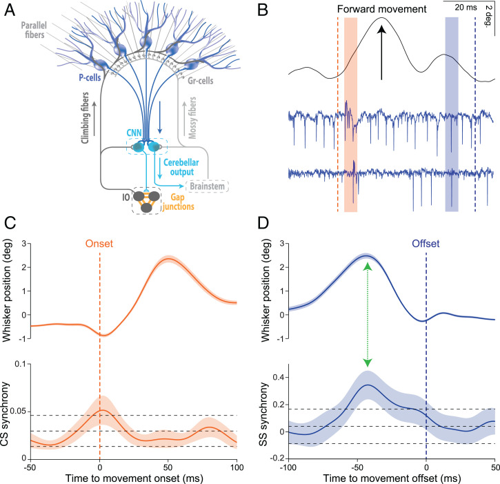
Synchrony of CSs and SSs may be correlated with onset and offset of movements, respectively. (A) Schematic representation of the convergence of inhibitory Purkinje cells (P-cells) onto cerebellar nuclei neurons (CNNs). P-cells integrate the inputs received via the climbing fiber and mossy fiber–parallel fiber systems and compute an output that can be read out by the CNNs. When the converging P-cell output is synchronous, the downstream effects at the CNNs may be subject to training and/or elicit sequences of relative silence and subsequent rebound activity. CNNs either directly control behavior via premotor areas in the brainstem or they provide feedback to neurons in the inferior olive (IO), which are coupled by gap junctions and provide the climbing fibers to the P-cells. Granular cells (Gr-cells) receive input from the mossy fibers and give rise to the parallel fibers. (B) Raw traces of a voluntary whisker movement (black line, top) and two simultaneously recorded PC-cells (blue traces, bottom). The orange and blue dashed lines indicate the onset and offset of the movement, respectively, while orange and blue beams indicate periods during which both PC-cells show CSs and SSs, respectively. (C) Average whisker movements (top) and synchrony of CS activity (bottom) centered around movement onset (dashed orange line). (D) Average whisker movements (top) and synchrony of SS activity (bottom) centered around movement offset (dashed blue line). CS synchrony rises just before movement onset and SS synchrony occurs before the whisker movements cease. Note that the peak of SS synchrony coincides at a moment of deceleration (green arrow), similar to the main finding by Sedaghat-Nejad et al. in PNAS (17). Top and bottom black dashed lines demarcate two SDs higher or lower than the mean of the synchrony levels as observed during the resting epoch (−200 to −150 ms prior to movement onset). CS and SS data are from 106 P-cell pairs from Romano et al. (18, 19). Note that whisker movements in B, C, and D are presented as position signals. Shaded areas indicate the SEM.
Comment on
-
Synchronous spiking of cerebellar Purkinje cells during control of movements.Sedaghat-Nejad E, Pi JS, Hage P, Fakharian MA, Shadmehr R. Sedaghat-Nejad E, et al. Proc Natl Acad Sci U S A. 2022 Apr 5;119(14):e2118954119. doi: 10.1073/pnas.2118954119. Epub 2022 Mar 29. Proc Natl Acad Sci U S A. 2022. PMID: 35349338 Free PMC article.
References
-
- Bell C. C., Grimm R. J., Discharge properties of Purkinje cells recorded on single and double microelectrodes. J. Neurophysiol. 32, 1044–1055 (1969). - PubMed
-
- Llinas R., Baker R., Sotelo C., Electrotonic coupling between neurons in cat inferior olive. J. Neurophysiol. 37, 560–571 (1974). - PubMed
-
- Sotelo C., Llinas R., Baker R., Structural study of inferior olivary nucleus of the cat: Morphological correlates of electrotonic coupling. J. Neurophysiol. 37, 541–559 (1974). - PubMed
Publication types
MeSH terms
LinkOut - more resources
Full Text Sources