This site needs JavaScript to work properly. Please enable it to take advantage of the complete set of features!
Skip to main page content
U.S. flag

An official website of the United States government

Dot gov

The .gov means it’s official.
Federal government websites often end in .gov or .mil. Before sharing sensitive information, make sure you’re on a federal government site.

Https

The site is secure.
The https:// ensures that you are connecting to the official website and that any information you provide is encrypted and transmitted securely.

NIH NLM Logo
Log in
Access keys NCBI Homepage MyNCBI Homepage Main Content Main Navigation
. 2022 Feb;100(2):620-637.
doi: 10.1002/jnr.24993. Epub 2021 Dec 1.

Input and output organization of the mesodiencephalic junction for cerebro-cerebellar communication

Affiliations

Input and output organization of the mesodiencephalic junction for cerebro-cerebellar communication

Xiaolu Wang et al. J Neurosci Res. 2022 Feb.

Abstract

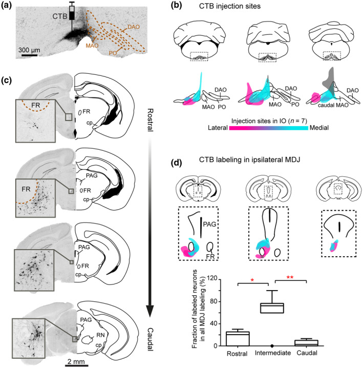
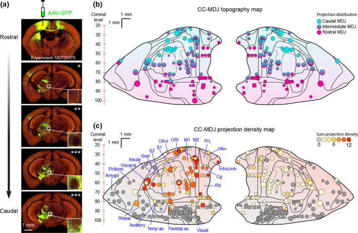
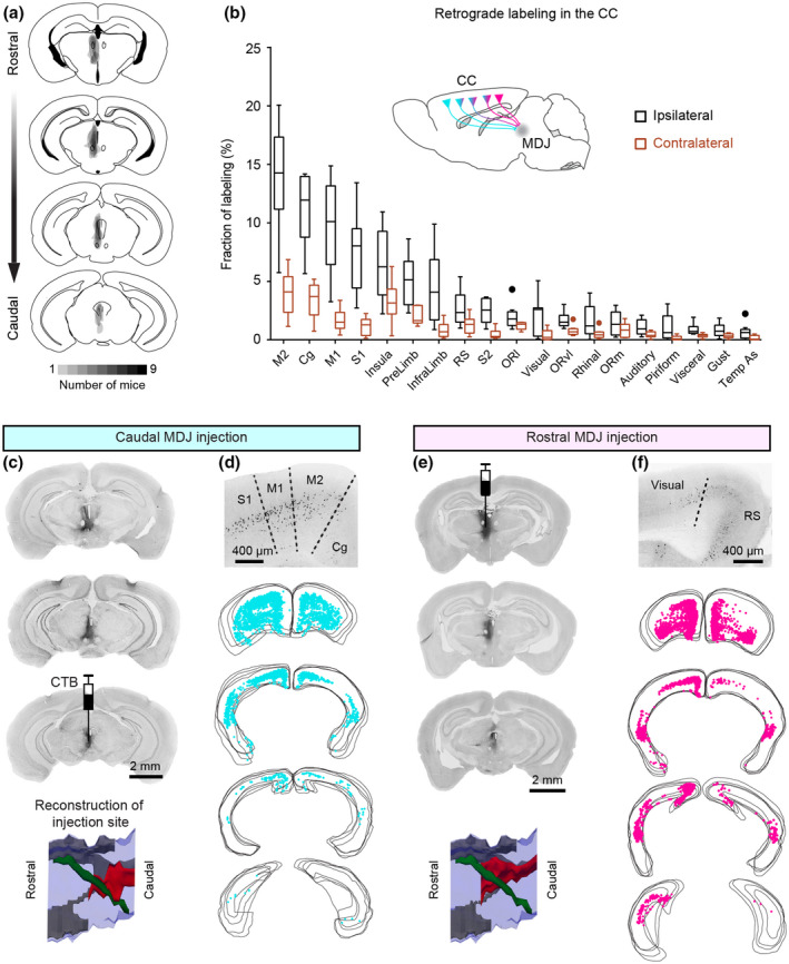
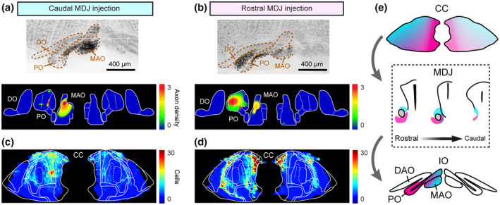
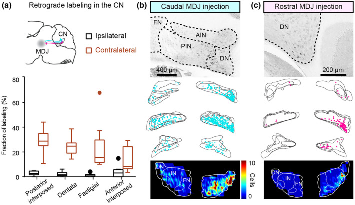
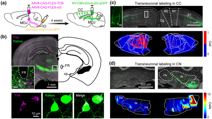
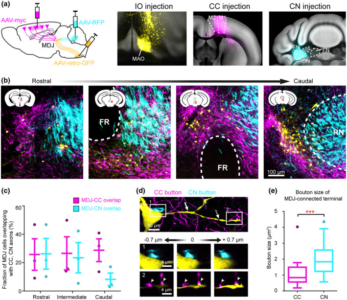
Most studies investigating the impact of the cerebral cortex (CC) onto the cerebellum highlight the role of the pons, which provides the mossy fibers to the cerebellum. However, cerebro-cerebellar communication may also be mediated by the nuclei of the mesodiencephalic junction (MDJ) that project to the inferior olive (IO), which in turn provides the climbing fibers to the molecular layer. Here, we uncover the precise topographic relations of the inputs and outputs of the MDJ using multiple, classical, and transneuronal tracing methods as well as analyses of mesoscale cortical injections from Allen Mouse Brain. We show that the caudal parts of the CC predominantly project to the principal olive via the rostral MDJ and that the rostral parts of the CC predominantly project to the rostral medial accessory olive via the caudal MDJ. Moreover, using triple viral tracing technology, we show that the cerebellar nuclei directly innervate the neurons in the MDJ that receive input from CC and project to the IO. By unraveling these topographic and prominent, mono- and disynaptic projections through the MDJ, this work establishes that cerebro-cerebellar communication is not only mediated by the pontine mossy fiber system, but also by the climbing fiber system.

Keywords: RRID:SCR_001622; RRID:SCR_002798; cerebellar nuclei; cerebral cortex; climbing fiber; inferior olive; mesodiencephalic junction (MDJ).


Conflict of interest statement

The authors declare no competing interests.

Figures

FIGURE 1
FIGURE 1
CTB injections in the IO retrogradely label MDJ neurons. (a) Coronal section of IO from a representative mouse, showing CTB injection in the MAO. (b) Summary of the IO injections for seven mice. Magenta, cyan, and gray: injections mainly cover PO (n = 2, magenta), rostral MAO (n = 3, cyan), and caudal MAO (n = 2, gray). (c) Retrogradely labeled neurons in the MDJ at four anteroposterior levels. Sections are obtained from the injection in (a). (d) Summary of CTB‐labeled neurons in the MDJ regions (upper, overlay of seven mice) and their anteroposterior distribution (lower). Error bars denote standard deviation. *p < 0.05; **p < 0.01
FIGURE 2
FIGURE 2
Viral anterograde tracing demonstrates CC‐MDJ topographic and density organizations. (a) Images of an example case from the Allen Mouse Brain Atlas with an injection in the motor cortex (top) and projections at anteroposterior levels of the MDJ. The number of asterisks represents cortical axon density in the MDJ (inset, scale bar = 100 μm, see quantification in the Materials and Methods). (b) CC‐MDJ topography map of cortical injections on our standard flattened CC representation (Aoki et al., 2019). Each marker represents one injection in the CC (wild types in circle, n = 83/167; transgenics in squares, n = 6/167); marker size indicates injection volume (see Materials and Method); color coding suggests axonal projection in the anteroposterior MDJ. (c) CC‐MDJ density map of cortical areas projecting onto the MDJ. The different density levels are represented by the color of the circles and squares on the standard flattened scheme of the CC. Note that the cases exhibiting no MDJ projection (gray markers, n = 78/167) were not illustrated in panel (b). Amygd, amygdala; Auditory, auditory cortex; Cg, cingulate cortex; Gust, gustatory cortex; InfraLimb, infralimbic cortex; Insula, insular cortex; M1, primary motor cortex; M2, secondary motor cortex; ORl, lateral orbital cortex; ORm, medial orbital cortex; ORvl, ventrolateral orbital cortex; Parietal as, parietal association cortex; Piriform, piriform cortex; PrL, prelimbic cortex; Rhinal, rhinal cortex; RS, retrosplenial cortex; S1, primary somatosensory cortex; S2, secondary somatosensory cortex; Temp as, temporal association cortex; Visceral, visceral cortex; Visual, visual cortex
FIGURE 3
FIGURE 3
CTB retrograde tracing from the MDJ to the CC. (a) Summary of CTB injections in the MDJ regions (n = 9 mice). (b) Quantification of retrogradely labeled cells in the CC. Light gray and dark gray represent fractions of total labeled cells on the contralateral and ipsilateral sides, respectively. (c–f) Comparison of retrograde cortical labeling from a caudal MDJ‐injected case and a rostral MDJ‐injected case. In the lateral view of injection site reconstruction (bottom, c and e), red: CTB injection site, green: FR, gray: third ventricle, blue: midbrain contour. Example cortical sections (d and f) show CTB labeling ipsilateral to the injection sites. In the serial plotting of cortical labeling (d and f), each panel include four consecutive sections. See abbreviations in Figure 2
FIGURE 4
FIGURE 4
CC‐MDJ‐IO topography by injecting CTB in anteroposterior MDJ. (a and b) Example sections (upper) and density maps (lower) of anterogradely labeled MDJ axons in the IO. (c and d) Density maps of retrogradely labeled neurons in the CC from the caudal and rostral MDJ injections. (e) Schematic of CC‐MDJ‐IO topographic organization. DO, dorsal accessory olive; MAO, medial accessory olive; PO, principal olive
FIGURE 5
FIGURE 5
Retrograde CTB labeling from the MDJ to the CN. (a) Summary of CTB‐labeled neurons in the CN (n = 9 mice). (b and c) Comparison of retrograde CN labeling from a caudal MDJ‐injected case and a rostral MDJ‐injected case. Example sections show labeling in the contralateral CN. In the serial plotting of CN labeling, each panel is overlayed with two to four consecutive sections. Note the variability of positional matching in the serial plotting is due to imperfect coronal sectioning. Labeling density is illustrated in the color maps (lower) for both cases
FIGURE 6
FIGURE 6
Monosynaptic rabies tracing revealing CC and CN inputs on the olivary‐projecting MDJ neurons. (a) Schematic of transneuronal viral‐tracing strategy (n = 4 mice). (b) Coronal sections of a representative mouse at the midbrain level (upper, inset suggesting MDJ, inset scale bar = 100 μm), showing rabies primary labeling (lower, TVA and rabies co‐labeled) and local secondary labeling. (c) Coronal section (upper) and plotting map (lower) of transneuronal rabies labeling in layer‐5 pyramidal cells (inset, scale bar = 50 μm) of motor cortex. (d) Same as (c), but for labeling in the CN. DN, dentate nucleus; FN, fastigial nucleus; IN, interposed nucleus
FIGURE 7
FIGURE 7
Convergence of CC and CN terminations on the olivary‐projecting MDJ neurons. (a) Schematic of tracing strategy. Coronal sections (registered in the Allen CCF) showing injections of retrograde AAV‐GFP in the IO, anterograde AAV‐myc in the motor cortex, and AAV‐RFP in the cerebellar interposed‐dentate complex. (b) Coronal sections of MDJ regions from an example mouse showing retrogradely labeled cells (olivary‐projecting cells, yellow) distributing in the CC (magenta) and CN (cyan) axons. (c) Colocalization quantification of olivary‐projecting cells and CC, CN axons at anteroposterior MDJ levels (n = 3 mice, plotted as mean ± SD). (d) Confocal images of an olivary‐projecting MDJ neuron from (b) showing close synaptic contact with CN axon on its soma (inset 1) and with CC axons on its secondary dendrite (inset 2). Solid arrow: primary dendrite, dashed‐line arrow: secondary dendrite. (e) Size comparison of CC and CN buttons exhibiting close contacts with IO‐projection MDJ cells (n = 27 for CC buttons, n = 37 for CN buttons, t (62) = 3.67, ***p < 0.001)

References

    1. Abdelgabar, A. R. , Suttrup, J. , Broersen, R. , Bhandari, R. , Picard, S. , Keysers, C. , De Zeeuw, C. I. , & Gazzola, V. (2019). Action perception recruits the cerebellum and is impaired in patients with spinocerebellar ataxia. Brain, 142, 3791–3805. 10.1093/brain/awz337 - DOI - PMC - PubMed
    1. Ackerley, R. , Pardoe, J. , & Apps, R. (2006). A novel site of synaptic relay for climbing fibre pathways relaying signals from the motor cortex to the cerebellar cortical C1 zone. Journal of Physiology, 576, 503–518. 10.1113/jphysiol.2006.114215 - DOI - PMC - PubMed
    1. Akkal, D. , Dum, R. P. , & Strick, P. L. (2007). Supplementary motor area and presupplementary motor area: Targets of basal ganglia and cerebellar output. Journal of Neuroscience, 27, 10659–10673. 10.1523/JNEUROSCI.3134-07.2007 - DOI - PMC - PubMed
    1. Amino, Y. , Kyuhou, S. , Matsuzaki, R. , & Gemba, H. (2001). Cerebello‐thalamo‐cortical projections to the posterior parietal cortex in the macaque monkey. Neuroscience Letters, 309, 29–32. 10.1016/S0304-3940(01)02018-3 - DOI - PubMed
    1. An, X. , Bandler, R. , Ongür, D. , & Price, J. L. (1998). Prefrontal cortical projections to longitudinal columns in the midbrain periaqueductal gray in macaque monkeys. Journal of Comparative Neurology, 401, 455–479. 10.1002/(SICI)1096-9861(19981130)401:4<455:AID-CNE3>3.0.CO;2-6 - DOI - PubMed

Publication types

Cite

AltStyle によって変換されたページ (->オリジナル) /