This site needs JavaScript to work properly. Please enable it to take advantage of the complete set of features!
Skip to main page content
U.S. flag

An official website of the United States government

Dot gov

The .gov means it’s official.
Federal government websites often end in .gov or .mil. Before sharing sensitive information, make sure you’re on a federal government site.

Https

The site is secure.
The https:// ensures that you are connecting to the official website and that any information you provide is encrypted and transmitted securely.

NIH NLM Logo
Log in
Access keys NCBI Homepage MyNCBI Homepage Main Content Main Navigation
. 2016 Jun 1;7(6):1069-1081.
doi: 10.1039/C6MD00005C. Epub 2016 Mar 28.

SLC Transporters: Structure, Function, and Drug Discovery

Affiliations

SLC Transporters: Structure, Function, and Drug Discovery

Claire Colas et al. Medchemcomm. .

Abstract

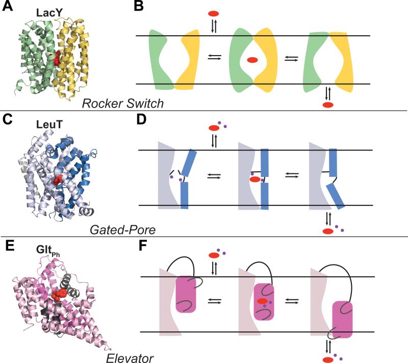
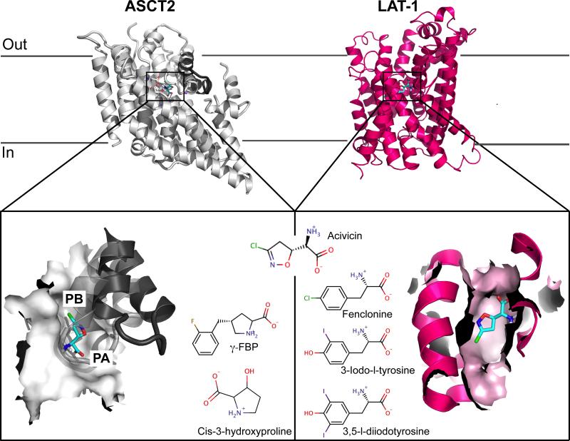
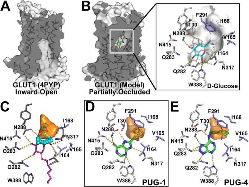
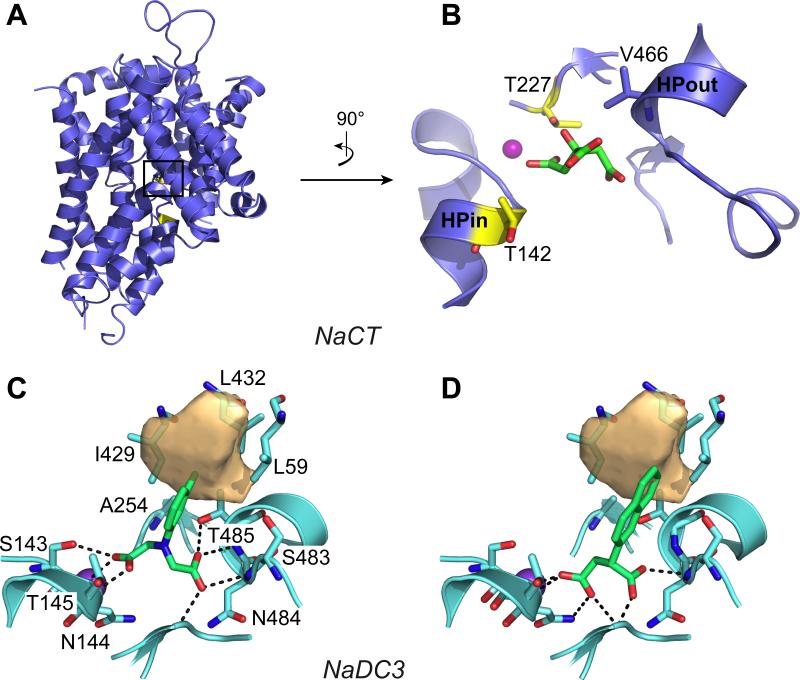
The human Solute Carrier (SLC) transporters are important targets for drug development. Structure-based drug discovery for SLC transporters requires the description of their structure, dynamics, and mechanism of interaction with small molecule ligands and ions. The recent determination of atomic structures of human SLC transporters and their homologs, combined with improved computational power and prediction methods have led to an increased applicability of structure-based drug design methods for human SLC members. In this review, we provide an overview of the SLC transporters' structures and transport mechanisms. We then describe computational techniques, such as homology modeling and virtual screening that are emerging as key tools to discover chemical probes for human SLC members. We illustrate the utility of these methods by presenting case studies in which rational integration of computation and experiment was used to characterize SLC members that transport key nutrients and metabolites, including the amino acid transporters LAT-1 and ASCT2, the SLC13 family of citric acid cycle intermediate transporters, and the glucose transporter GLUT1. We conclude with a brief discussion about future directions in structure-based drug discovery for the human SLC superfamily, one of the most structurally and functionally diverse protein families in human.


Figures

Figure 1
Figure 1. Transport mechanism models
In each case, a representative structure of a transporter that uses the mechanism described is shown in (A, C, E) and three states of the transport cycle of the associated transporter are depicted in (B, D, F): outward-open, occluded and inward-open. Rocker Switch. (A) LacY is shown in an outward-open bound conformation (PDB: 4OAA) with the N- and C-terminal halves in green and yellow cartoons respectively, and the substrate in red spheres. (B) The substrate binds to a V-shape conformation facing the extracellular side of the membrane, triggering an intermediate occluded state. The substrate is then released from an inverted V-shape inward-open conformation. Gated-pore. (C) LeuT is shown an outward-open bound conformation (PDB: 4FXZ), with the scaffold and bundle domains represented in light and dark blue cartoons respectively, the substrate and ions in red and purple spheres. (D) The scaffold domain remains static, whereas the bundle domain experiences conformational changes to bind and release the substrate. The binding site is enclosed by a thin gate (i.e., a salt bridge) on the extracellular side, and a thick gate (TM1) on the intracellular side. Elevator. (E) GltPh is shown in an outward-open bound conformation (PDB: 2NWW), with the oligomerization and transport domain represented in light pink and magenta cartoons, respectively, and the two gates (HP1 and HP2) in gray cartoons. The inhibitor is shown in red spheres, and sodium ions in purple spheres. (F) The oligomerization domain remains invariant while the transport domain moves in a piston-like movement to transport the substrate from the extracellular side to the intracellular side of the membrane.
Figure 2
Figure 2. Homology models of the amino acid transporters ASCT2 and LAT-1 bound to acivicin
The top panel shows both transporters viewed from the membrane bilayer plane. ASCT2 is represented in gray cartoons, with the HP2 gate in the outward-open conformation shown in dark gray cartoons. LAT-1 is represented in pink cartoons. The bottom panels show the binding site of each transporter, represented in surface, with acivicin docked. Pockets A and B revealed by the models of ASCT2 are labeled PA and PB, respectively. The newly discovered ligands are shown next to their respective targets.
Figure 3
Figure 3. Structure-based drug discovery targeting the occluded conformation of the human GLUT1
Comparison of GLUT1 in (A) crystal structure in an inward-open conformation (PDBID: 4PYP) and (B) homology model in an occluded conformation with the glucose-binding site highlighted in surface representation. (C) The H-pocket, lined by hydrophobic residues (blue sticks), observed in the occluded model (orange volume) is larger than in the inward-open structure (cyan spheres), e.g. 4PYP (grey sticks and magenta alkyl sugar analog). (D and E) Ligands (green sticks) discovered through virtual screening against the occluded GLUT1 model (white sticks) occupy the glucose-binding site and the H-pocket (orange volume).
Figure 4
Figure 4. Homology models of the human transporter NaCT (SLC13A5) and NaDC3 (SLC13A3)
(A) The NaCT model in an inward-open conformation is shown in blue cartoon within the membrane plane. The NaCT binding site, with citrate (green sticks) and Na+ (purple sphere) is shown in (B). The position of the Na+ is derived from the X-ray structure of the template structure VcINDY; the coordinates of citrate are predicted with docking. The two binding site residues (yellow sticks) whose mutations are associated with epileptic encephalopathy are shown. The predicted binding pose of two of the newly discovered SLC13 ligands (green sticks) in NaDC3 are shown in (C) and (D). The NaDC3 binding site is represented in cyan cartoons, with the residues interacting with the ligands in sticks. The hydrophobic pocket specific to this transporter is shown in light orange volume.

References

    1. Schlessinger A, Khuri N, Giacomini KM, Sali A. Curr Top Med Chem. 2013;13:843–856. - PMC - PubMed
    1. Lin L, Yee SW, Kim RB, Giacomini KM. Nature reviews. Drug discovery. 2015;14:543–560. - PMC - PubMed
    1. Cesar-Razquin A, Snijder B, Frappier-Brinton T, Isserlin R, Gyimesi G, Bai X, Reithmeier RA, Hepworth D, Hediger MA, Edwards AM, Superti-Furga G. Cell. 2015;162:478–487. - PubMed
    1. Bareford LM, Swaan PW. Advanced drug delivery reviews. 2007;59:748–758. - PMC - PubMed
    1. Chang C, Ekins S, Bahadduri P, Swaan PW. Adv Drug Deliv Rev. 2006;58:1431–1450. - PMC - PubMed
Cite

AltStyle によって変換されたページ (->オリジナル) /