This site needs JavaScript to work properly. Please enable it to take advantage of the complete set of features!
Skip to main page content
U.S. flag

An official website of the United States government

Dot gov

The .gov means it’s official.
Federal government websites often end in .gov or .mil. Before sharing sensitive information, make sure you’re on a federal government site.

Https

The site is secure.
The https:// ensures that you are connecting to the official website and that any information you provide is encrypted and transmitted securely.

NIH NLM Logo
Log in
Access keys NCBI Homepage MyNCBI Homepage Main Content Main Navigation
Review
. 2013 Dec 27:7:123.
doi: 10.3389/fnsys.2013.00123.

Developmental plasticity of spatial hearing following asymmetric hearing loss: context-dependent cue integration and its clinical implications

Affiliations
Review

Developmental plasticity of spatial hearing following asymmetric hearing loss: context-dependent cue integration and its clinical implications

Peter Keating et al. Front Syst Neurosci. .

Abstract

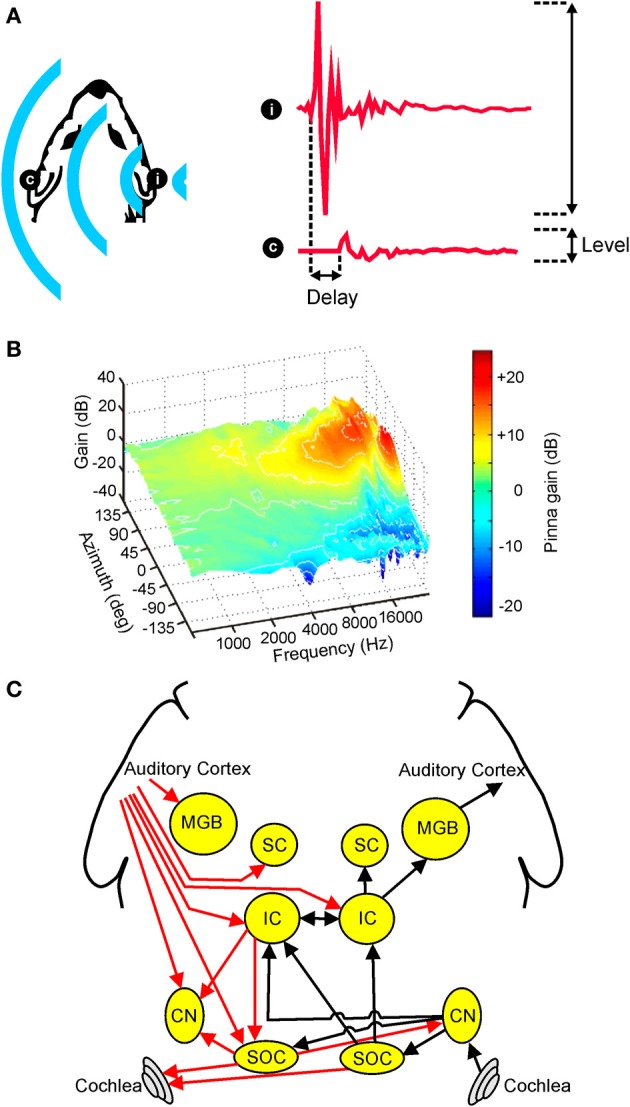
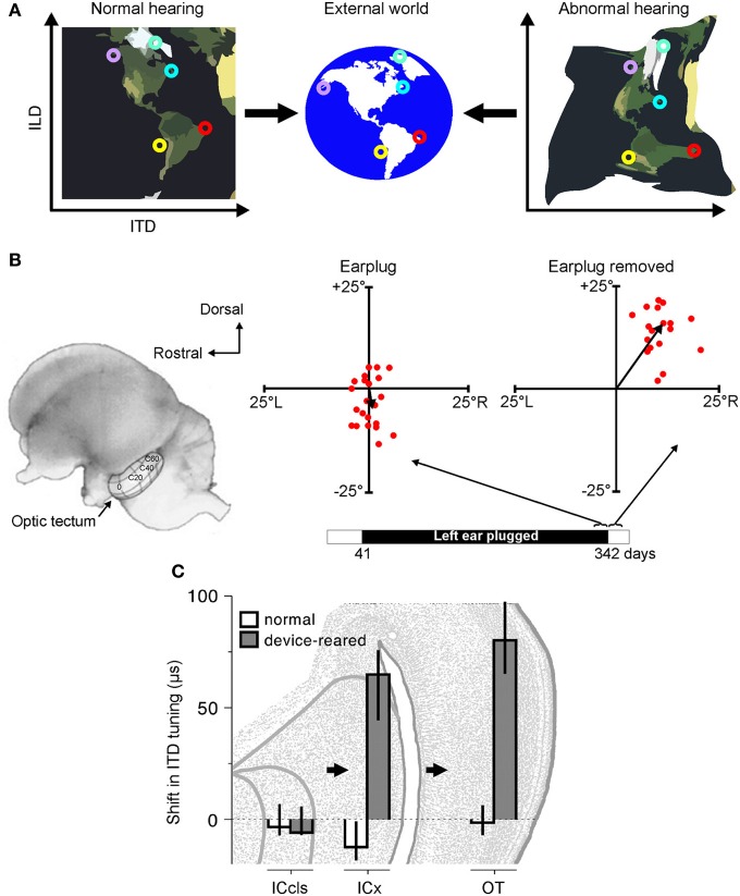
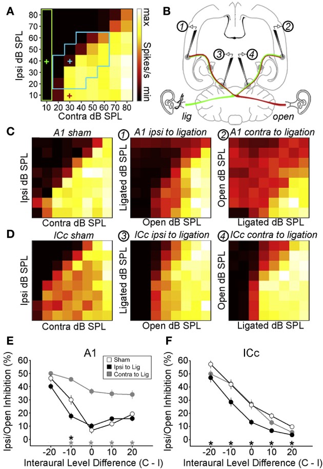
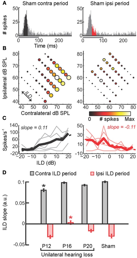
Under normal hearing conditions, comparisons of the sounds reaching each ear are critical for accurate sound localization. Asymmetric hearing loss should therefore degrade spatial hearing and has become an important experimental tool for probing the plasticity of the auditory system, both during development and adulthood. In clinical populations, hearing loss affecting one ear more than the other is commonly associated with otitis media with effusion, a disorder experienced by approximately 80% of children before the age of two. Asymmetric hearing may also arise in other clinical situations, such as after unilateral cochlear implantation. Here, we consider the role played by spatial cue integration in sound localization under normal acoustical conditions. We then review evidence for adaptive changes in spatial hearing following a developmental hearing loss in one ear, and show that adaptation may be achieved either by learning a new relationship between the altered cues and directions in space or by changing the way different cues are integrated in the brain. We next consider developmental plasticity as a source of vulnerability, describing maladaptive effects of asymmetric hearing loss that persist even when normal hearing is provided. We also examine the extent to which the consequences of asymmetric hearing loss depend upon its timing and duration. Although much of the experimental literature has focused on the effects of a stable unilateral hearing loss, some of the most common hearing impairments experienced by children tend to fluctuate over time. We therefore propose that there is a need to bridge this gap by investigating the effects of recurring hearing loss during development, and outline recent steps in this direction. We conclude by arguing that this work points toward a more nuanced view of developmental plasticity, in which plasticity may be selectively expressed in response to specific sensory contexts, and consider the clinical implications of this.

Keywords: adaptation; auditory localization; binaural; conductive hearing loss; cortex; learning; midbrain; monaural.


Figures

Figure 1
Figure 1
Overview of auditory spatial processing. (A) Sample acoustic waveforms are shown for the ipsilateral (i) and contralateral (c) ears following presentation of sound from a source located to one side of the head. The incoming sound is typically delayed and attenuated in the contralateral ear, producing interaural time differences (ITDs) and interaural level differences (ILDs), respectively. These cues are referred to as binaural spatial cues as they depend on comparisons between the two ears. (B) Gain is plotted for an adult ferret ear as a function of sound frequency and azimuth. Because the filtering effects of the head and ears depend on the direction of a sound source, the observed spectra vary with respect to azimuth (and elevation), producing spectral shape cues to sound location. (C) Simplified schematic of the mammalian auditory pathway showing the principal ascending (black) and descending (red) projections between the cochlea, cochlear nuclei (CN), superior olivary complex (SOC), inferior colliculus (IC), superior colliculus (SC), medial geniculate body (MGB), and auditory cortex. For clarity, each of these projections is shown for one side of the brain only. (B) adapted with permission from King et al. (2001).
Figure 2
Figure 2
Adaptation to abnormal cues can be achieved by remapping the relationship between cue values and sound-source location. (A) Under normal listening conditions (left), specific combinations of cue values correspond to particular locations in the external world. Small circles of the same color represent particular cue combinations and their corresponding locations in the external world. Under abnormal listening conditions, such as when one ear is occluded by an earplug, these relationships are distorted and altered (right). In order to use these abnormal cues for accurate sound localization, the brain must therefore learn that the same locations now correspond to different cue combinations. (B) At present, robust neurophysiological evidence for cue remapping has only been observed in barn owls reared with one ear occluded. Electrophysiological recordings from neurons in the optic tectum of these animals show that compensatory shifts take place in the neurons' auditory spatial tuning. Tectal neurons respond most strongly to visual and auditory stimuli presented from overlapping locations, with their receptive fields arranged systematically to produce topographically-aligned maps of visual and auditory space (represented by the contour lines superimposed on the optic tectum in the picture of the owl's brain). Recordings from the rostral region of the tectum in an owl that was reared with the left ear occluded until 342 days after hatching revealed little difference between the visual and auditory receptive field centers when the earplug was still in place ("Earplug"). Misalignment in elevation is plotted on the ordinate, with misalignment in azimuth plotted on the abscissa. When the ear was occluded, the data points cluster around the origin, demonstrating that auditory and visual receptive fields are broadly in register. Following earplug removal, however, the receptive fields became systematically misaligned in both azimuth and elevation, indicating that the neurons were tuned to binaural cue values that no longer corresponded to their preferred visual location. (C) Site of auditory plasticity in the ascending auditory pathway of the barn owl. Frequency-dependent shifts in ITD tuning are plotted for barn owls reared either with normal hearing or with a passive filtering device in one ear that delays and attenuates sound. Positive values indicate shifts in ITD tuning that compensate for the effects of the device. Bars and lines show medians and interquartile ranges. Data are shown for the optic tectum (OT), external nucleus of the inferior colliculus (ICx) and the lateral shell of the central nucleus of the inferior colliculus (ICcls). Shifts in ITD tuning emerge at the level of the ICx. Modified with permission from Knudsen (1985) and Gold and Knudsen (2000b).
Figure 3
Figure 3
Adapting to a unilateral hearing loss by changing the dependence of the auditory system on different spatial cues. (A) Performance on an approach-to-target sound localization task is shown for normally-reared, control ferrets fitted with an earplug in one ear for the first time, as well as ferrets reared with a unilateral earplug (juvenile-plugged) and tested with an earplug in the developmentally-occluded ear. The animals initiated a trial by waiting on a central platform and approached the source of a sound presented from one of 12 loudspeakers positioned at equal intervals around the periphery, as illustrated in the accompanying schematic (top right). Juvenile-plugged animals performed the task with much greater accuracy than controls. (B) When spatial cues are altered or degraded by hearing loss, the auditory system can adapt by becoming less dependent on the abnormal cues and more dependent on the cues that remain intact. (C) Cue reweighting in primary auditory cortex. Neural weighting index is shown for neurons in the primary auditory cortex of juvenile-plugged ferrets while a virtual earplug was experienced in the developmentally-occluded ear. Stimuli were presented over earphones so that individual cues could be manipulated independently, which enabled a weighting index to be constructed. Higher values indicate that relatively more weight was given to the spatial cues provided by the intact ear. Data are also shown for normally-reared, control animals experiencing a virtual earplug in one ear. Neural weighting index values are higher in juvenile-plugged animals than controls, indicating greater reliance on the unchanged spectral cues provided by the intact ear. (D–F) Behavioral reweighting of auditory spatial cues revealed using reverse correlation. If approach to target localization responses are determined by the spectral cues provided by the intact ear, it is possible to recover these cues using reverse correlation. Juvenile-plugged ferrets performed the task in (A), but the stimulus spectra were randomized across trials. The mean stimulus spectrum across all trials was very close to zero (gray line). However, on the subset of trials on which behavioral responses were made to a particular location (60° in the example shown), the mean stimulus spectrum deviated considerably from zero, with distinct spectral features emerging at frequencies >16 kHz (D). Repeating this analysis for each response location produced a reverse correlation map (E), which closely resembled the directional transfer function (DTF) of the intact (right) ear (F). These results indicate that localization behavior in juvenile-plugged animals is guided by spectral features that resemble those produced by the directional filtering properties of the intact ear. This was not the case in controls, indicating that the juvenile-plugged animals had developed a greater dependence on, and therefore adapted to the unilateral hearing loss by giving greater weight to, the spectral cues that are unaffected by an earplug. Modified with permission from Keating et al. (2013a).
Figure 4
Figure 4
Maladaptive effects of asymmetric hearing loss during development. (A) Example of a binaural interaction matrix recorded from a unit in the central nucleus of the inferior colliculus (ICc) in a normally reared rat. Contralateral sound level is plotted against ipsilateral level, with color denoting the number of spikes fired for each combination. Firing rates typically increase as the contralateral level is increased, but are suppressed when the ipsilateral level exceeds that in the contralateral ear. For each interaural level combination enclosed by the blue box, binaural suppression was quantified by comparing it with the linear sum of its monaural intercepts (e.g., blue cross relative to the sum of the red and green crosses). (B) To investigate the developmental effects of monaural deprivation, rats were reared with a hearing loss in one ear that was induced by ligation of the ear canal, which was reversed prior to electrophysiological experiments. Bilateral recordings were then performed in the ICc and primary auditory cortex (A1) of these animals and compared with data from sham-operated controls reared with normal hearing. (C,D) Examples of binaural interaction matrices from A1 (C) and ICc (D) in sham operated controls (left), and in ligated animals. For ligated animals, data are shown for the hemisphere ipsilateral (middle) and contralateral (right) to the ligated ear. Color scales and axis labels are identical to (A). (E,F) Ipsilaterally mediated suppression expressed as a function of ILD for A1 (E) and ICc (F) recordings. Data are shown for sham operated controls (open symbols) as well as ligated animals, both contralateral (gray) and ipsilateral (black) to the ligated ear. Asterisks denote significant differences between ligated animals and controls, with asterisk grayscale indicating the hemisphere in which the comparison was made. Error bars show SEMs. Ipsilaterally mediated suppression in A1 of monaurally deprived animals is increased in the hemisphere contralateral to the deprived ear (E), but not in the corresponding hemisphere of the ICc (F). Conversely, ipsilaterally mediated suppression is reduced in the ICc ipsilateral to the deprived ear (F), but this effect is not apparent at the level of A1 (E). These results suggest that monaural deprivation induces persistent changes in the strength of ipsilateral input, which acts to weaken the representation of the deprived ear at the level of the ICc and strengthen the representation of the intact ear at the level of A1. In both cases, this increases the relative strength of the intact ear, and produces maladaptive shifts in ILD sensitivity. Modified with permission from Popescu and Polley (2010).
Figure 5
Figure 5
Precise timing of unilateral hearing loss influences developmental outcome. (A) Poststimulus time histograms (PSTHs) of spikes recorded from neurons in the primary auditory cortex of the mouse can be divided into windows that show the strongest differential sensitivity to contralateral (black, left) and ipsilateral (red, right) ILDs. Greater contralateral ILD sensitivity is typically a feature of short-latency responses, whereas greater sensitivity to ipsilateral ILDs tends to be seen in longer latency responses. (B) Firing rate as a function of ILD at 5 different average binaural levels, with each plot corresponding to the response window shown immediately above. Heat map is scaled to the normalized firing rate within each time window, whereas circle diameter is normalized to the maximum firing rate across both time windows. (C) Faint lines show firing rates as a function of ILD for each of the average binaural levels shown in (B). Thicker lines show the average of ILD functions obtained with different average binaural levels. (D) Slopes of linear fits were calculated for the thick lines in (C) and provide a measure of ILD sensitivity, with larger slope values indicating greater sensitivity. Mean slope values (±s.e.m.) are shown for sham operated controls as well as mice that experienced brief periods (1–2 weeks) of unilateral hearing loss beginning at different ages (either postnatal day 12, 16, or 20). Contralateral and ipsilateral ILD sensitivity are both reduced by asymmetric hearing loss, but these different aspects of spatial processing are vulnerable at different stages of development. Asterisk indicates significant differences relative to (sham-operated) controls (post-hoc tests following ANOVA, P < 0.05). Modified with permission from Polley et al. (2013).
Figure 6
Figure 6
Otitis media with effusion (OME) and its effects in humans. (A) State transition matrix for different effusion states, obtained via a prospective study among a group of 95 children aged 0–3 years. Arrow color shows the probability of transitioning from one effusion state to another. Effusion was either present (filled circles) or absent (open circles) in the left (L) and right (R) ears. Individuals often alternate between periods of normal and abnormal hearing. (B) Masking level difference, which provides a measure of spatial listening in noisy environments, is shown for adults as well as children with a history of OME (mean ± s.e.m.). Children are divided into quartiles depending on the amount of OME experienced during the first 5 years of life, with the colors indicating the mean prevalence of OME in each quartile. Adults with unknown OME experience are shown in gray. A deficit in masking level difference was observed in the upper quartile of children that experienced OME, corresponding to children that experienced OME at least 50% of the time. Based on Hogan et al. (1997) and Hogan and Moore (2003).
Figure 7
Figure 7
Context-dependent reweighting of auditory spatial cues following a recurring developmental hearing loss in one ear. (A) Sound localization performance of ferrets reared with an earplug in one ear, either in the presence or absence of an earplug. Each symbol represents data from an individual animal. Although juvenile-plugged ferrets adapt to an earplug (see Figure 3), their performance improves when the earplug is removed, and approaches the mean performance level of controls under normal listening conditions (dotted black line). This means that juvenile-plugged ferrets adapt to an asymmetric hearing loss without compromising their ability to localize accurately when normal hearing becomes available. Error bars denote bootstrapped 95% confidence intervals, with solid black lines showing group means. (B) Context-specific reweighting of auditory spatial cues. Randomizing stimulus spectra across trials is known to degrade the usefulness of spectral cues, since it becomes unclear whether spectral features arise from the filtering effects of the head and ears or are instead properties of the stimulus itself. Whilst wearing an earplug, sound localization performance in juvenile-plugged ferrets declined as the amount of spectral randomization was increased, but this effect largely disappeared once the earplug was removed. Each line shows data for an individual animal, either with an earplug in place (solid, dark blue), following earplug removal (solid, pink), or after the reintroduction of an earplug (dotted, dark blue). (C) To quantify the effects of spectral randomization, slope values were calculated for the lines in (B) and are plotted for different hearing conditions. Each symbol shows data from an individual animal, with solid black lines indicating group means. Performance of juvenile-plugged ferrets was only impaired by randomization (negative slope values) when one ear was occluded. This means that the localization behavior of juvenile-plugged ferrets became more dependent on the spectral cues available to the contralateral ear whenever a unilateral earplug was present. The lack of effect of spectral randomization in the absence of the earplug suggests that the animals were relying on binaural cues when normal inputs were available. (D) Neural weighting index values are shown for neurons in the primary auditory cortex of juvenile-plugged ferrets, either in the presence or absence of a virtual earplug. Higher values indicate greater reliance on the spectral cues provided by the developmentally non-occluded ear. In juvenile-plugged ferrets, neural weighting index values change depending on whether a virtual earplug is present or not. Controls do not show the same effect, and are indistinguishable from juvenile-plugged ferrets under normal hearing conditions (dotted black line shows mean neural weighting index values for controls). This means that recurring monaural deprivation during development leads to cortical neurons weighting auditory spatial cues differently depending on whether a hearing loss is experienced, providing a possible neural basis for the cue reweighting observed behaviorally. Modified with permission from Keating et al. (2013a).

References

    1. Agterberg M. J., Hol M. K., Cremers C. W., Mylanus E. A., Van Opstal J., Snik A. F. (2011). Conductive hearing loss and bone conduction devices: restored binaural hearing? Adv. Otorhinolaryngol. 71, 84–91 10.1159/000323587 - DOI - PubMed
    1. Agterberg M. J., Snik A. F., Hol M. K., Van Wanrooij M. M., Van Opstal A. J. (2012). Contribution of monaural and binaural cues to sound localization in listeners with acquired unilateral conductive hearing loss: improved directional hearing with a bone-conduction device. Hear. Res. 286, 9–18 10.1016/j.heares.2012年02月01日2 - DOI - PubMed
    1. Alais D., Burr D. (2004). The ventriloquist effect results from near-optimal bimodal integration. Curr. Biol. 14, 257–262 10.1016/S0960-9822(04)00043-0 - DOI - PubMed
    1. Alais D., Newell F. N., Mamassian P. (2010). Multisensory processing in review: from physiology to behaviour. Seeing Perceiving 23, 3–38 10.1163/187847510X488603 - DOI - PubMed
    1. Alvarado J. C., Stanford T. R., Vaughan J. W., Stein B. E. (2007). Cortex mediates multisensory but not unisensory integration in superior colliculus. J. Neurosci. 27, 12775–12786 10.1523/JNEUROSCI.3524-07.2007 - DOI - PMC - PubMed

LinkOut - more resources

Cite

AltStyle によって変換されたページ (->オリジナル) /