This site needs JavaScript to work properly. Please enable it to take advantage of the complete set of features!
Skip to main page content
U.S. flag

An official website of the United States government

Dot gov

The .gov means it’s official.
Federal government websites often end in .gov or .mil. Before sharing sensitive information, make sure you’re on a federal government site.

Https

The site is secure.
The https:// ensures that you are connecting to the official website and that any information you provide is encrypted and transmitted securely.

NIH NLM Logo
Log in
Access keys NCBI Homepage MyNCBI Homepage Main Content Main Navigation
. 2009 Dec 8;106(49):20752-7.
doi: 10.1073/pnas.0908570106. Epub 2009 Nov 19.

Inward-facing conformation of glutamate transporters as revealed by their inverted-topology structural repeats

Affiliations

Inward-facing conformation of glutamate transporters as revealed by their inverted-topology structural repeats

Thomas J Crisman et al. Proc Natl Acad Sci U S A. .

Abstract

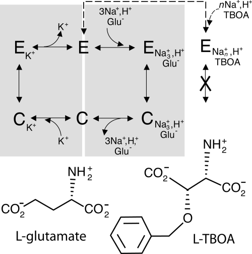
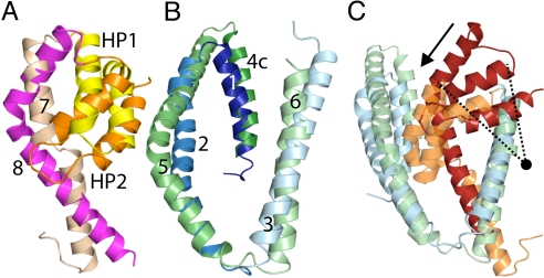
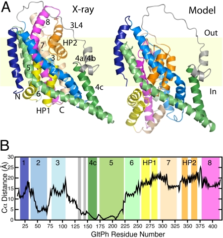
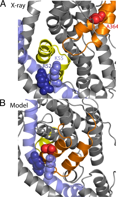
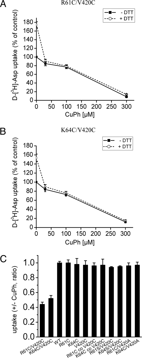
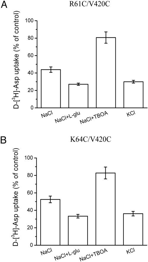
Glutamate transporters regulate synaptic concentrations of this neurotransmitter by coupling its flux to that of sodium and other cations. Available crystal structures of an archeal homologue of these transporters, GltPh, resemble an extracellular-facing state, in which the bound substrate is occluded only by a small helical hairpin segment called HP2. However, a pathway to the cytoplasmic side of the membrane is not clearly apparent. We previously modeled an alternate state of a transporter from the neurotransmitter:sodium symporter family, which has an entirely different fold, solely on the presence of inverted-topology structural repeats. In GltPh, we identified two distinct sets of inverted-topology repeats and used these repeats to model an inward-facing conformation of the protein. To test this model, we introduced pairs of cysteines into the neuronal glutamate transporter EAAC1, at positions that are >27 A apart in the crystal structures of GltPh, but approximately = 10 A apart in the inward-facing model. Transport by these mutants was activated by pretreatment with the reducing agent dithithreitol. Subsequent treatment with the oxidizing agent copper(II)(1,10-phenantroline)(3) abolished this activation. The inhibition of transport was potentiated under conditions thought to promote the inward-facing conformation of the transporter. By contrast, the inhibition was reduced in the presence of the nontransportable substrate analogue D,L-threo-beta-benzyloxyaspartate, which favors the outward-facing conformation. Other conformation-sensitive accessibility measurements are also accommodated by our inward-facing model. These results suggest that the inclusion of inverted-topology repeats in transporters may provide a general solution to the requirement for two symmetry-related states in a single protein.


Conflict of interest statement

The authors declare no conflict of interest.

Figures

Fig. 1.
Fig. 1.
Transport by the mammalian EAATs. The transport cycle of mammalian EAATs involves two half-cycles (gray boxes). First, sodium, protons, and glutamate from the extracellular medium bind to an extracellular-facing conformation of the protein (E). A subsequent conformation change to form a cytoplasm-facing conformation (C) allows access to the binding site from the cytoplasm so that the substrates can be released. In the second half-cycle, intracellular potassium binds, the protein returns to an extracellular-facing conformation, and potassium can be released to the extracellular medium. Thus, addition of potassium to the extracellular solution increases the proportion of transporters in an inward-facing conformation. Transport can be interrupted by binding of nontransportable glutamate analogues (TBOA) with sodium, locking the protein in an outward-facing conformation.
Fig. 2.
Fig. 2.
Structural similarity and differences between segments of GltPh. Structural alignments of (A) HP1 (yellow) plus TM7 (pale orange), against HP2 (orange) plus TM8 (magenta), and (B) TM 1 to 3 (shades of blue) with TM 4c to 6 (shades of green). In (C) we considered TM 1 to 3 (blue), plus HP2 and TM8 (red) as one pseudorepeat, and TM 4c-6 (green), HP1 and TM7 (orange) as a second repeat. Note that only the first three TMs were used for the fit: this structural superposition highlights the shift in position of the hairpin-TM motif with respect to the first three TMs of each pseudorepeat (arrow), which is a vertical curving movement (the approximate pivot point is shown as a circle). Figures were made using PyMOL (33).
Fig. 3.
Fig. 3.
Organization of repeats in GltPh. (A) Schematic of the topology of GltPh emphasizing the structural relationships between the four segments, I to IV. (B) Schematic of the sequence alignment used to generate the swapped-repeat model. For example, segment I was modeled using the structure of segment II as a template. The conformation of the loop between TMs 3 and 4 (3L4) was taken from the x-ray structure.
Fig. 4.
Fig. 4.
Model of GltPh in a cytoplasm-facing conformation. (A) Structure of a protomer of the cytoplasm-facing model (Right) compared with the x-ray crystal structure of the extracellular-facing conformation (Left). The protein is viewed along the plane of the membrane, with the extracellular side at the top, and colored as in Fig. 3. (B) Distance between Cα atoms in the outward-facing structure and the model of the inward-facing conformation. For this calculation, the structures were superposed using TMs 4c and 5 (residues 151–218) at the subunit interface, which is believed not to change during transport (14).
Fig. 5.
Fig. 5.
Cross-linking residues become close in the cytoplasm-facing model of GltPh. Residues R52 (dark blue spheres), K55 (blue spheres), and A364 (red spheres) from TM2 (blue) and HP2 (orange) are shown in the x-ray structure (A) and in the cytoplasm-facing model (B). These residues correspond, respectively, to R61C, K64C, and V420C of EAAC1.
Fig. 6.
Fig. 6.
Effect of DTT and CuPh on d-[3H]aspartate transport by R61C/V420C and K64C/V420C. Transport of d-[3H]aspartate was measured in Xenopus laevis oocytes expressing the indicated mutants after pretreatment for 5 min in the presence or absence of 5-mM DTT, followed by a 5-min incubation with CuPh, as described in Materials and Methods. (A and B) Initial treatment in the presence or absence of 5-mM DTT was followed by a 5-min incubation with the indicated concentrations of CuPh. Results are expressed as percent of activity of oocytes that were preincubated twice for 5 min in frog Ringer's solution without DTT or CuPh. (C) After preincubation with DTT, the oocytes were incubated for 5 min in the presence or absence of 100-μM CuPh. The results are expressed as the ratio of uptake in the presence of CuPh over that in its absence. "R61C co V420C" and "K64C co V420C" represent oocytes in which the two indicated cRNAs were coinjected.
Fig. 7.
Fig. 7.
Effect of the medium composition during CuPh treatment on the transport activity of R61C/V420C and K64C/V420C. Oocytes expressing R61C/V420C (A) or K64C/V420C (B) were pretreated with 5-mM DTT for 5 min, followed by incubation for 5 min with 100-μM CuPh in either: frog Ringer's solution in the absence or presence of either 1-mM l-glutamate or 60 μM of TBOA or frog Ringer's solution in which all of the NaCl was replaced by KCl. The results are expressed as a percentage of activity of oocytes incubated without CuPh.

References

    1. Zerangue N, Kavanaugh MP. Flux coupling in a neuronal glutamate transporter. Nature. 1996;383:634–637. - PubMed
    1. Levy LM, Warr O, Attwell D. Stoichiometry of the glial glutamate transporter GLT-1 expressed inducibly in a chinese hamster ovary cell line selected for low endogenous Na+-dependent glutamate uptake. J Neurosci. 1998;18:9620–9628. - PMC - PubMed
    1. Kanner BI, Bendahan A. Binding order of substrates to the sodium and potassium ion coupled l-glutamic acid transporter from rat brain. Biochem. 1982;21:6327–6330. - PubMed
    1. Pines G, Kanner BI. Counterflow of l-glutamate in plasma membrane vesicles and reconstituted preparations from rat brain. Biochem. 1990;29:11209–11214. - PubMed
    1. Kavanaugh MP, Bendahan A, Zerangue N, Zhang Y, Kanner BI. Mutation of an amino acid residue influencing potassium coupling in the glutamate transporter GLT-1 induces obligate exchange. J Biol Chem. 1997;272:1703–1708. - PubMed

Publication types

MeSH terms

Cite

AltStyle によって変換されたページ (->オリジナル) /