This site needs JavaScript to work properly. Please enable it to take advantage of the complete set of features!
Skip to main page content
U.S. flag

An official website of the United States government

Dot gov

The .gov means it’s official.
Federal government websites often end in .gov or .mil. Before sharing sensitive information, make sure you’re on a federal government site.

Https

The site is secure.
The https:// ensures that you are connecting to the official website and that any information you provide is encrypted and transmitted securely.

NIH NLM Logo
Log in
Access keys NCBI Homepage MyNCBI Homepage Main Content Main Navigation
. 2009 May 1;182(9):5702-11.
doi: 10.4049/jimmunol.0803450.

IL-7 receptor expression provides the potential for long-term survival of both CD62Lhigh central memory T cells and Th1 effector cells during Leishmania major infection

Affiliations

IL-7 receptor expression provides the potential for long-term survival of both CD62Lhigh central memory T cells and Th1 effector cells during Leishmania major infection

Sara L Colpitts et al. J Immunol. .

Abstract

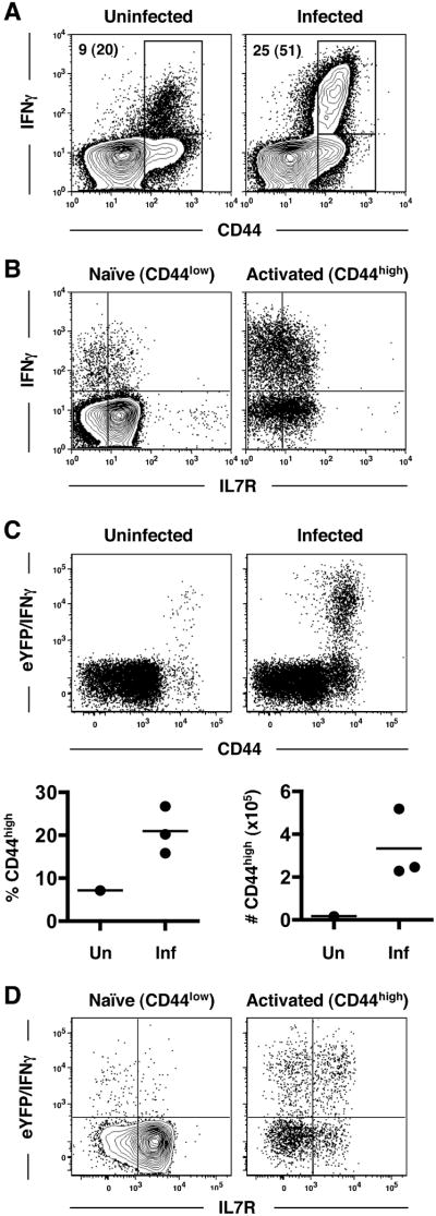
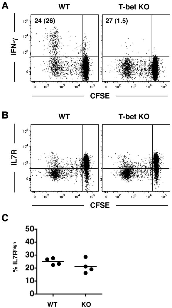
Infection with the intracellular protozoan parasite Leishmania major induces a state of concomitant immunity wherein secondary immunity is dependent upon the persistence of the original pathogen. Our laboratory has described two populations of Leishmania-induced CD4(+) T cells that contribute to immunity: CD62L(high) central memory T (T(CM)) cells and CD62L(low) effector T cells. To determine whether the prosurvival cytokine IL-7 contributes to maintaining these T cells, we examined expression of the IL7R on CD4(+) T cells activated during L. major infection. We found that T(CM) cells present in chronically infected mice expressed high levels of the IL7R. However, in addition to the expression of the IL7R by T(CM) cells, CD62L(low) cells responding to L. major infection expressed the IL7R. Additional experiments revealed that a large percentage of the IL7R(high)CD62L(low) cells were Th1 cells, based on transcription at the IFN-gamma locus and up-regulation of the Th1-promoting transcription factor T-bet. The up-regulation of T-bet did not prevent IL7R expression by L. major-responding CD4(+) T cells, nor did the absence of T-bet result in increased IL7R expression. Finally, blockade of IL7R signaling decreased the number of T-bet(+)CD4(+) T cells, reduced IFN-gamma production, and inhibited delayed-type hypersensitivity responses in immune mice challenged with L. major, indicating that IL7R signaling contributes to the maintenance of Th1 effector cells. Thus, both T(CM) and Th1 effector cells can express the IL7R during chronic L. major infection, which provides a potential means for their long-term survival in addition to the presence of persisting parasites.


Conflict of interest statement

Disclosures The authors have no financial conflict of interest.

Figures

Figure 1
Figure 1. CD62Lhigh TCM cells generated in response to L. major infection express high levels of the IL7R
CD4+ T cells were enriched using MACS columns from the lymphoid tissue of C57BL/6 mice (CD45.2) that had been infected in the footpad with L. major 12 weeks prior to harvest. The cells were CFSE-labeled and transferred to naïve CD45.1 recipients. Recipient mice were subsequently infected in the ear with L. major. Control mice were left uninfected for the duration of the experiment. Cells were isolated from the auricular draining lymph node (dLN) after 14 days. Plots have been gated on the CD4+CD45.2+CD45.1- lymphocytes and are representative of 8 mice in 3 separate experiments. IL7R expression (black line) on the CFSEbriCD62Lhigh cells of uninfected controls (top) and CFSEdimCD62Lhigh cells of infected mice (bottom) is shown relative to isotype controls (shaded histogram). (B) CD4+CD62LhighIL7Rhigh cells from naïve or immune mice (>12 weeks pi) were sorted on a FACS Aria with purity of >80%. The cells were CFSE-labeled, and 1 ×ばつ 106 cells were transferred to congenic recipients as above one day prior to infection with L. major. Plots are gated on CD45.2+CD45.1- lymphocytes and represent 5-6 mice per group in 2 independent experiments. Numbers indicate the percent CFSEdim of the donor cell populations. (C) Cytokine production by the donor cells from infected mice in B was assessed by intracellular cytokine staining. Numbers represent the percent cytokine positive of the CFSEdim donor population.
Figure 2
Figure 2. The population of activated CD4+ T cells only transiently downregulates IL7R expression revealing 2 populations of IL7R-expressing cells during the primary immune response to infection
(A) C57BL/6 mice were infected in the footpad with L. major, and at the indicated weeks pi, the draining popliteal lymph node was isolated for flow cytometry. Plots have been gated on CD4+ cells, and the relative levels of IL7R expression are shown on CD44low (shaded histograms) and CD44high (black line) cells. (B) CD62L expression is shown on the CD44high IL7Rhigh cells (black line) in A. CD62L expression on the naïve CD44low IL7Rhigh cells (shaded histograms) is shown for comparison. Data are representative of 6 mice in 2 independent experiments.
Figure 3
Figure 3. Th1-polarized cells can express the IL7R
(A) C57BL/6 mice were infected in the footpad with L. major. After 2 weeks, the draining popliteal LN was isolated from infected mice, and peripheral LNs were also isolated from uninfected controls. Lymphocytes were examined for IFN-γ production by intracellular cytokine staining. Plots have been gated on CD4+ T cells. Numbers indicate the percentage of CD4+ T cells that are CD44high with the percentage of CD44high cells producing IFN-γ in parenthesis. (B) IL7R expression versus IFN-γ production is shown in the CD44low versus CD44high cells from the infected mouse in A. (C) Spleens and lymph nodes were isolated from Yeti mice (CD45.2) and stained for CD4 and CD44 prior to cell sorting. Naïve CD45.1 recipient mice received 4 ×ばつ 106 CD4+CD44loweYFPneg cells, were infected in the ear with L. major, and then sacrificed after 2 weeks. Plots have been gated on CD45.2+CD45.1- donor cells from the auricular dLN of infected (inf) mice and uninfected (un) controls. (D) IL7R expression versus eYFP/IFN-γ production is shown on donor CD4+ cells from infected mice in C on the Leishmania-induced CD44high cells and naïve controls (CD44low). Plots are representative of 3 (un) and 7 (inf) mice in 2 independent experiments.
Figure 4
Figure 4. A population of IL7R-expressing cells has also upregulated T-bet expression
CD4+ T cells were MACS-purified from either naïve (A) or immune (B) donor mice prior to CFSE-labeling, transferred to naïve congenic recipients, and infected in the ear with L. major. After 2 weeks, dLNs (auricular) were isolated for flow cytometry. Plots have been gated on the donor CD4+CD45.2+CD45.1- cells. Additional plots indicate expression of CD62L, IL7R, and T-bet on the CFSEdim cells. Data are representative of at least 5 mice in 3 independent experiments. In C and D, naïve CFSE-labeled donor cells were transferred to naïve congenic recipients as in A, and IL-12 was administered with the parasites at the time of infection and additionally on d3 and d7 in the indicated mice. Numbers in C represent to percent IL7Rhigh T-betpos of the CD62Llow population ± SEM. Asterisks (*) indicate significance of p < 0.05.
Figure 5
Figure 5. Leishmania-responding CD4+ T cells express similar levels of the IL7R in the absence of T-bet expression
CD4+ T cells were MACS-purified from naïve WT or T-bet KO donors. Cells were CFSE-labeled and transferred to naïve CD45.1 congenic recipients that were infected in the ear with L. major the following day. At 2 weeks pi, auricular dLNs were analyzed for CFSE dilution and (A) IFN-γ production or (B) IL7R expression. Plots are gated on CD45.2+CD45.1- cells and are representative of 8 mice in 2 independent experiments. Numbers in A indicate the percentage of donor cells that are CFSEdim with the percent IFN-γpos of the CFSEdim population in parenthesis. The percentage of CFSEdim cells expressing the IL7R is shown in C, which was not significant (n.s.) between the 2 groups.
Figure 6
Figure 6. CD4+ effector T cells utilize IL-7 in immune mice
Immune C57BL/6 mice were either treated with blocking antibody against the IL7R (A7R34; 200μg every 2-3 days) or left untreated as controls. After 2 weeks of treatment, the animals were sacrificed, and the total numbers of cells in each of the indicated populations were calculated (A-C). Data are presented as the mean ± the SEM. Splenocytes were also stimulated with freeze-thawed leishmanial antigen (FTAg), and the supernatants collected after 72hr of culture were assayed for IFN-γ production (D). A second group of Ab-treated mice was challenged with L. major in the contralateral footpad, and DTH was measured by footpad swelling at 48 hours post secondary challenge (E) Parasite burden was determined by limiting dilution assay 2 weeks after challenge (F) during which time no anti-IL7R treatment was administered. DTH and parasite burden in naïve mice are shown as a control. Asterisks (*) indicate significance of p < 0.05.

References

    1. Sacks D, Noben-Trauth N. The immunology of susceptibility and resistance to Leishmania major in mice. Nat Rev Immunol. 2002;2:845–858. - PubMed
    1. Sypek JP, Chung CL, Mayor SE, Subramanyam JM, Goldman SJ, Sieburth DS, Wolf SF, Schaub RG. Resolution of cutaneous leishmaniasis: interleukin 12 initiates a protective T helper type 1 immune response. J Exp Med. 1993;177:1797–1802. - PMC - PubMed
    1. Heinzel FP, Schoenhaut DS, Rerko RM, Rosser LE, Gately MK. Recombinant interleukin 12 cures mice infected with Leishmania major. J Exp Med. 1993;177:1505–1509. - PMC - PubMed
    1. Nadim A, Javadian E, Tahvildar-Bidruni G, Ghorbani M. Effectiveness of leishmanization in the control of cutaneous leishmaniasis. Bull Soc Pathol Exot Filiales. 1983;76:377–383. - PubMed
    1. Scott P, Artis D, Uzonna J, Zaph C. The development of effector and memory T cells in cutaneous leishmaniasis: the implications for vaccine development. Immunol Rev. 2004;201:318–338. - PubMed

Publication types

MeSH terms

Cite

AltStyle によって変換されたページ (->オリジナル) /