This site needs JavaScript to work properly. Please enable it to take advantage of the complete set of features!
Skip to main page content
U.S. flag

An official website of the United States government

Dot gov

The .gov means it’s official.
Federal government websites often end in .gov or .mil. Before sharing sensitive information, make sure you’re on a federal government site.

Https

The site is secure.
The https:// ensures that you are connecting to the official website and that any information you provide is encrypted and transmitted securely.

NIH NLM Logo
Log in
Access keys NCBI Homepage MyNCBI Homepage Main Content Main Navigation
. 2002 Feb 15;22(4):1454-67.
doi: 10.1523/JNEUROSCI.22-04-01454.2002.

The coding of spatial location by single units in the lateral superior olive of the cat. I. Spatial receptive fields in azimuth

Affiliations

The coding of spatial location by single units in the lateral superior olive of the cat. I. Spatial receptive fields in azimuth

Daniel J Tollin et al. J Neurosci. .

Abstract

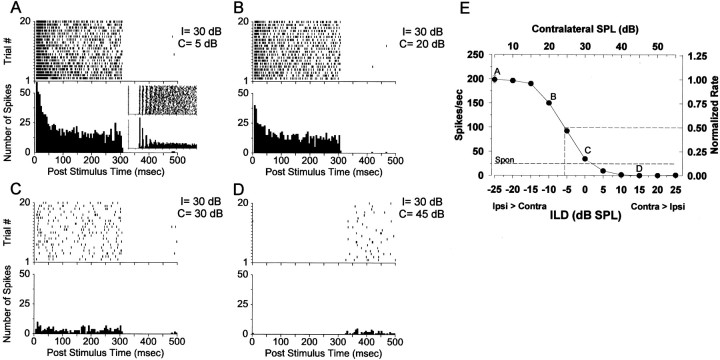
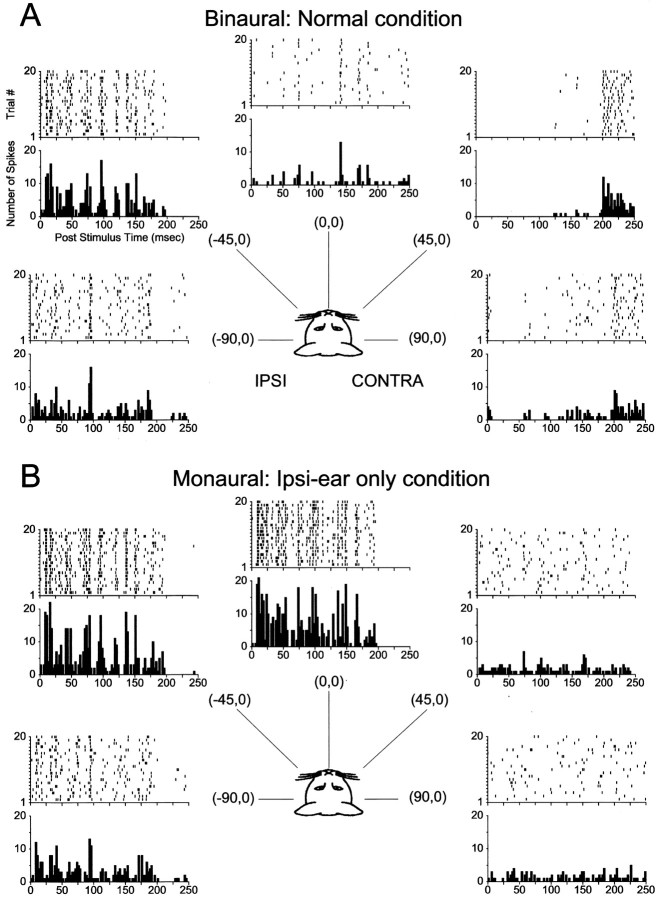
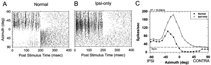
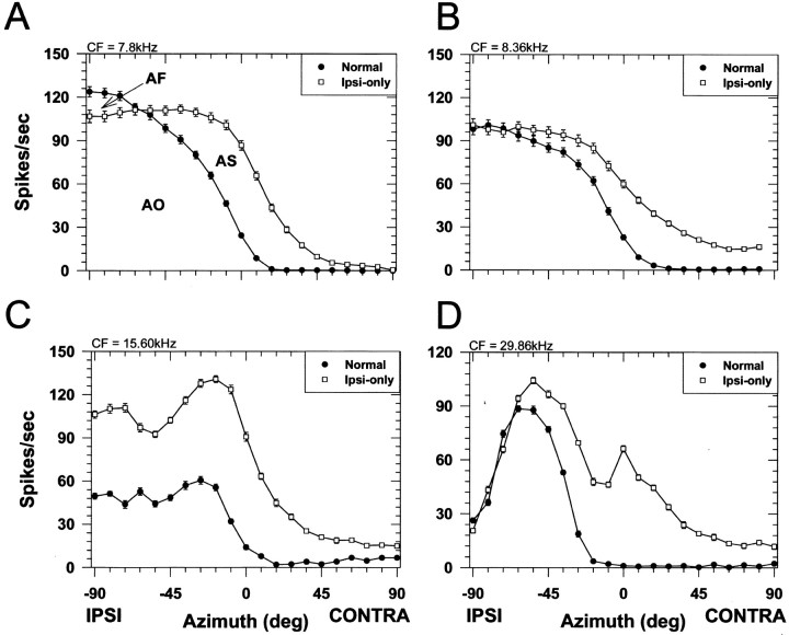
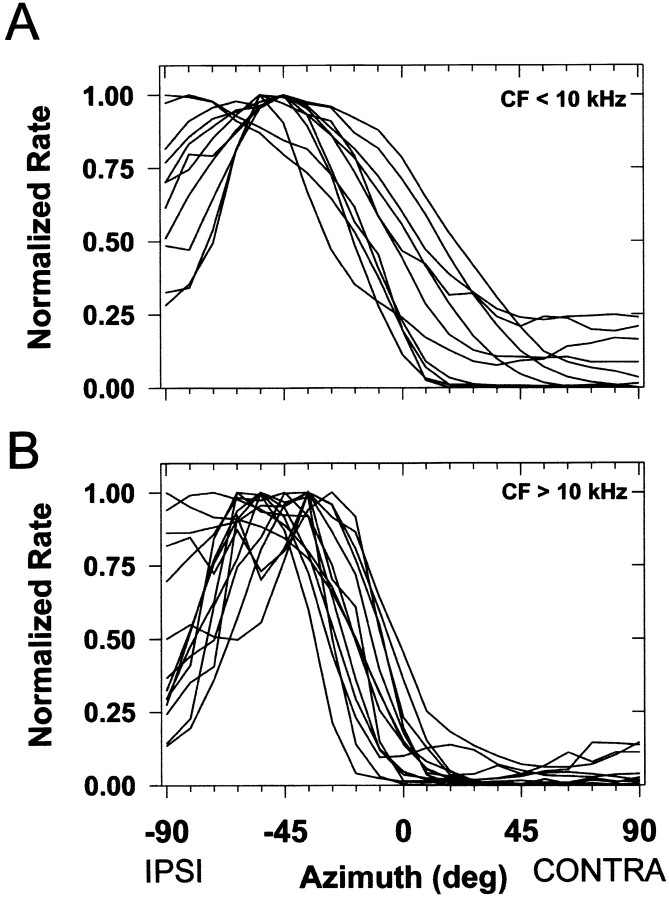
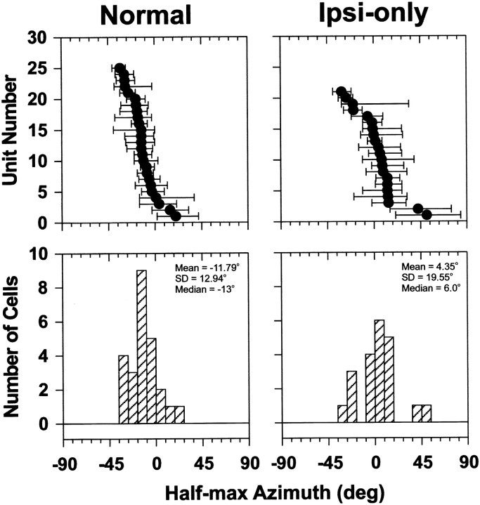
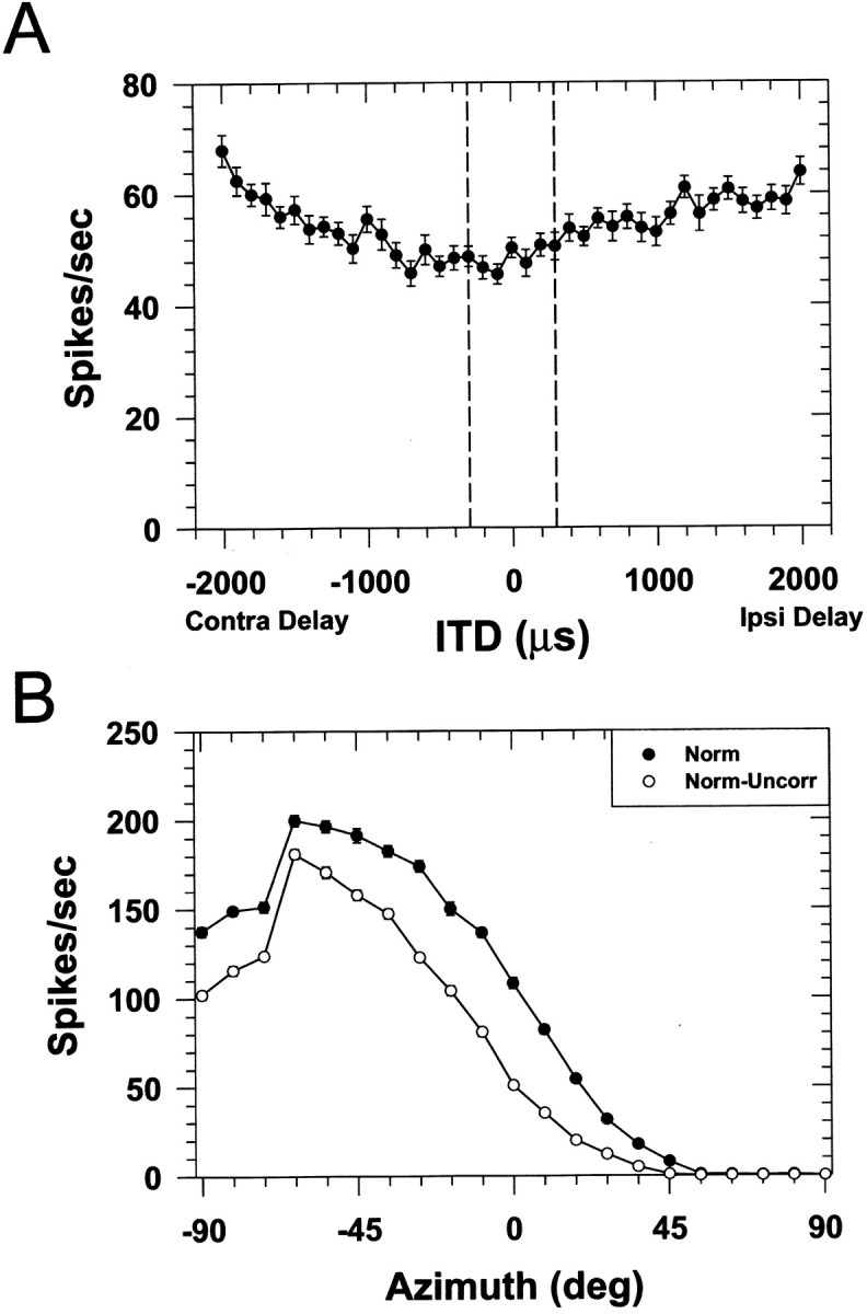
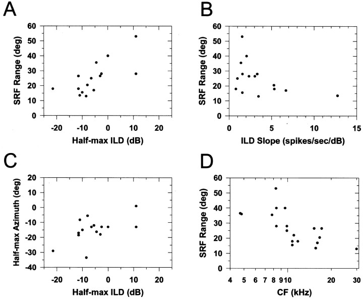
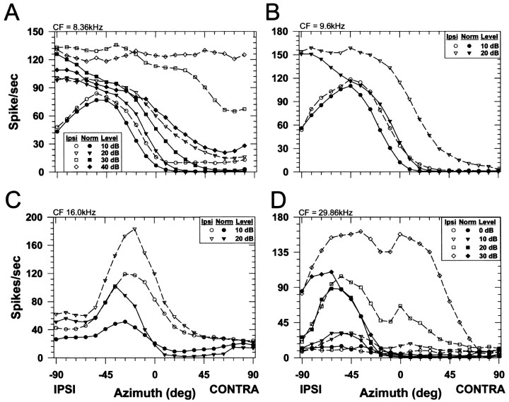
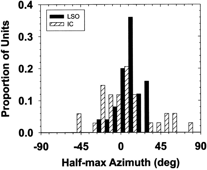
The lateral superior olive (LSO) is one of the most peripheral auditory nuclei receiving inputs from both ears, and LSO neurons are sensitive to interaural level differences (ILDs), one of the primary acoustical cues for sound location. We used the virtual space (VS) technique to present over earphones broadband stimuli containing natural combinations of localization cues as a function of azimuth while recording extracellular responses from single LSO cells. The responses of LSO cells exhibited spatial receptive fields (SRFs) in azimuth consonant with their sensitivity to ILDs of stimuli presented dichotically: high discharge rates for ipsilateral azimuths where stimulus amplitude to the excitatory ear exceeded that to the inhibitory ear, rapidly declining rates near the midline, and low rates for contralateral azimuths where the amplitude to the inhibitory ear exceeded that to the excitatory ear. Relative to binaural stimulation, presentations of the VS stimuli to the ipsilateral ear alone yielded increased rates, particularly in the contralateral field, confirming that the binaural SRFs were shaped by contralateral inhibition. Our finding that LSO neurons respond to azimuth consistent with their ILD sensitivity supports the long-held hypothesis that LSO neurons compute a correlate of the ILD present in free-field stimuli. Only weak correlations between the properties of pure-tone ILD functions and the SRFs were found, indicating that ILD sensitivity measured at only one sound level is not sufficient to predict sensitivity to azimuth. Sensitivity to spatial location was also retained over a wide range of stimulus levels under binaural, but not monaural, conditions.


Figures

Fig. 1.
Fig. 1.
Responses of an LSO cell to variations in the ILD of a CF tone. A–D, Dot rasters and PST histograms in response to a 300 msec ipsilateral tone at 30 dB SPL as a function of the level of a tone at the contralateral ear, as shown in the top right of each panel. Theinset in A shows the first 40 msec of the response to short tone pips at CF presented monaurally to the ipsilateral ear only, demonstrating the characteristic chopping response exhibited by most of our cells (bin width, 400 μsec and the top tic on the ordinate, 150 spikes). E, Mean discharge rate ± 1 SEM versus ILD (in decibels: SPL). (In this and all subsequent figures, where the error bars are not present, the SEM is less than the height of the data point.) The top abscissaindicates the level of the tone at the contralateral ear, and theright ordinate shows the rate normalized to the maximum. Thedashed horizontal line shows the spontaneous rate of the unit.
Fig. 2.
Fig. 2.
Responses of the same cell in Figure 1 to 200 msec broadband noise presented from five different azimuths along the horizontal plane under binaural (A, normal) and monaural ipsilateral-ear only (B, ipsi-only) conditions. Negative azimuths indicate sound sources in the ipsilateral sound field. The stimulus was presented at ∼20 dB above threshold at (0°,0°) in the ipsi-only condition. The top andbottom figures in each panel show the dot rasters and PST histograms for 20 presentations of the stimuli at each azimuth.
Fig. 3.
Fig. 3.
A,B, Temporal discharge patterns for the same cell in Figure 2 plotted continuously over the 20 stimulus presentations as a function of each of 21 azimuths for the normal (A) and ipsi-only (B) conditions. Each tic mark on theordinate of A and B denotes a different azimuthal position. C, The spatial receptive fields in azimuth for the ipsi-only and normal stimulus conditions. Each data point plots the mean discharge rate of the cell averaged over the 200 msec duration of the noise stimuli and 20 presentations. Thehorizontal line indicates the spontaneous rate.
Fig. 4.
Fig. 4.
Spatial receptive fields of four LSO cells. The CF of each cell is indicated in the top left of each figure. Same format as in Figure 3C.
Fig. 5.
Fig. 5.
Spatial receptive field shape depends on CF.A, Normalized SRFs for LSO cells with CFs <10 kHz.B, Normalized SRFs for cells with CFs >10 kHz.
Fig. 6.
Fig. 6.
The top panels plot the distribution of half-maximal azimuths (filled circles) and the ranges of azimuth corresponding to 25–75% of maximum discharge (bars). Data for the normal and ipsi-only conditions are shown in the left andright columns, respectively. Note that the data for each cell is plotted as a function of increasing half-maximal azimuth toward the ipsilateral sound field for both the normal and ipsi-only configurations. As a result, the same cell number in each plot does not necessarily designate the same cell. The bottom panelsshow histograms of the normal and ipsi-only half-maximal azimuths. The normal binaural spatial receptive fields are located predominantly in the ipsilateral sound field, whereas the ipsi-only receptive fields are located more into the contralateral field.
Fig. 7.
Fig. 7.
Plot of the BIT versus the BIS for all LSO units. The dotted lines indicate the boundaries suggested byDelgutte et al. (1999) to classify units into four separate categories of binaural interaction: monaural (Mon.), binaural facilitation (BF), binaural inhibition (BI), and mixed facilitatory–inhibitory interactions (BF&I).
Fig. 8.
Fig. 8.
A, Responses of one LSO cell to broadband noise as a function of the interaural time difference. Data points plot the mean discharge rate ± 1 SEM. Negative delays indicate that the onset of the noise to the contralateral ear was delayed with respect to that at the ipsilateral ear. Thevertical dashed lines indicate the range of ITDs expected for the average adult cat. The response of the cell is not modulated by ongoing ITDs in broadband noise over this range of ITDs.B, Normal binaural SRFs of another LSO cell under conditions in which the broadband noises presented to the two ears were identical (filled circles) or uncorrelated (open circles).
Fig. 9.
Fig. 9.
Scatterplots of the properties of the azimuthal SRFs and the properties of the ILD functions measured with tones at CF.A, The SRF range versus the half-maximal ILD.B, The SRF range versus the slope of the ILD function.C, The half-maximal azimuth of the SRFs versus the half-maximal ILD. D, The SRF range versus the CF of the unit.
Fig. 10.
Fig. 10.
The effect of overall sound level on the SRFs of four LSO cells. Open and filled symbolsindicate the ipsi-only and normal stimulus conditions, respectively. The parameter is the overall stimulus level above the threshold level for the ipsi-only condition measured at (0°, 0°). The SEM is not shown in this figure.
Fig. 11.
Fig. 11.
Histogram of half-maximal azimuths of SRFs measured using similar stimuli in LSO and the 34 IC cells from the study of Delgutte et al. (1999) exhibiting predominantly EI binaural interaction. The half-maximal azimuths of the LSO cells have been reflected about the midline.

References

    1. Adam TJ, Schwarz DW, Finlayson PG. Firing properties of chopper and delay neurons in the lateral superior olive of the rat. Exp Brain Res. 1999;124:489–502. - PubMed
    1. Adams JC, Mugnaini E. Dorsal nucleus of the lateral lemniscus: a nucleus of GABAergic projection neurons. Brain Res Bull. 1984;13:585–590. - PubMed
    1. Aitkin L, Jones R. Azimuthal processing in the posterior auditory thalamus of cats. Neurosci Lett. 1992;142:81–84. - PubMed
    1. Aitkin LM, Martin RL. The representation of stimulus azimuth by high best-frequency azimuth-selective neurons in the central nucleus of the inferior colliculus of the cat. J Neurophysiol. 1987;57:1185–1200. - PubMed
    1. Altshuler MW, Comalli PE. Effect of stimulus intensity and frequency on median horizontal plane sound localization. J Aud Res. 1975;15:262–265.

Publication types

LinkOut - more resources

Cite

AltStyle によって変換されたページ (->オリジナル) /