This site needs JavaScript to work properly. Please enable it to take advantage of the complete set of features!
Skip to main page content
U.S. flag

An official website of the United States government

Dot gov

The .gov means it’s official.
Federal government websites often end in .gov or .mil. Before sharing sensitive information, make sure you’re on a federal government site.

Https

The site is secure.
The https:// ensures that you are connecting to the official website and that any information you provide is encrypted and transmitted securely.

NIH NLM Logo
Log in
Access keys NCBI Homepage MyNCBI Homepage Main Content Main Navigation
. 2000 Dec 1;529 Pt 2(Pt 2):505-15.
doi: 10.1111/j.1469-7793.2000.00505.x.

Sensory integration in the perception of movements at the human metacarpophalangeal joint

Affiliations

Sensory integration in the perception of movements at the human metacarpophalangeal joint

D F Collins et al. J Physiol. .

Abstract

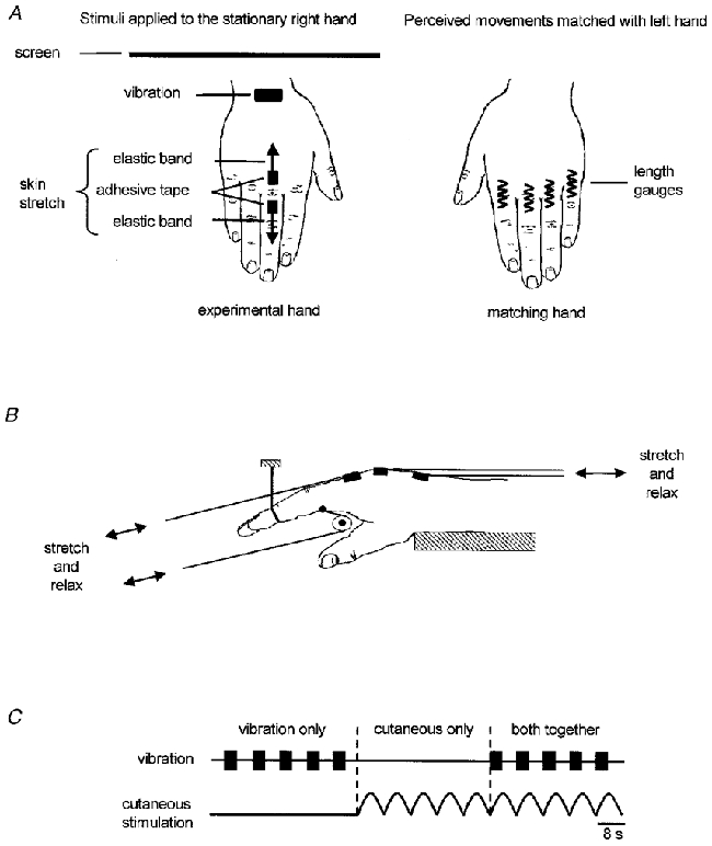
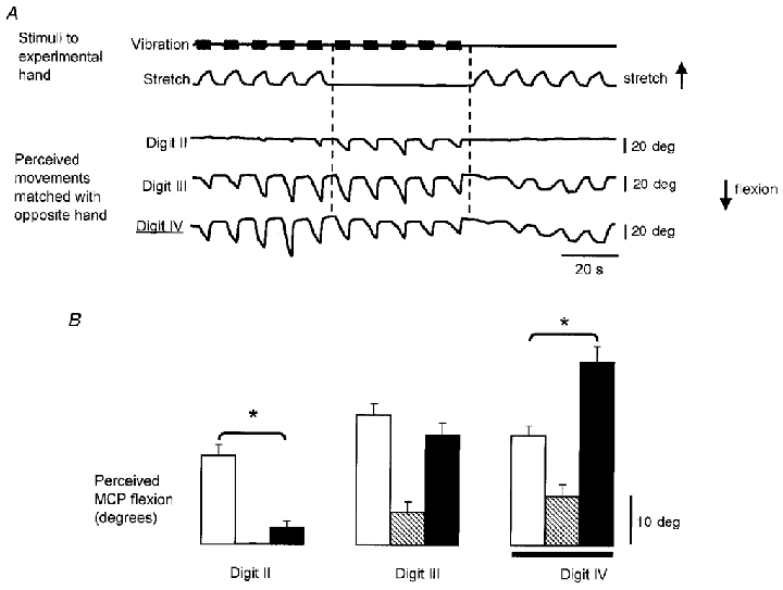
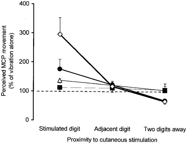
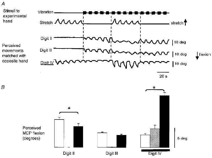
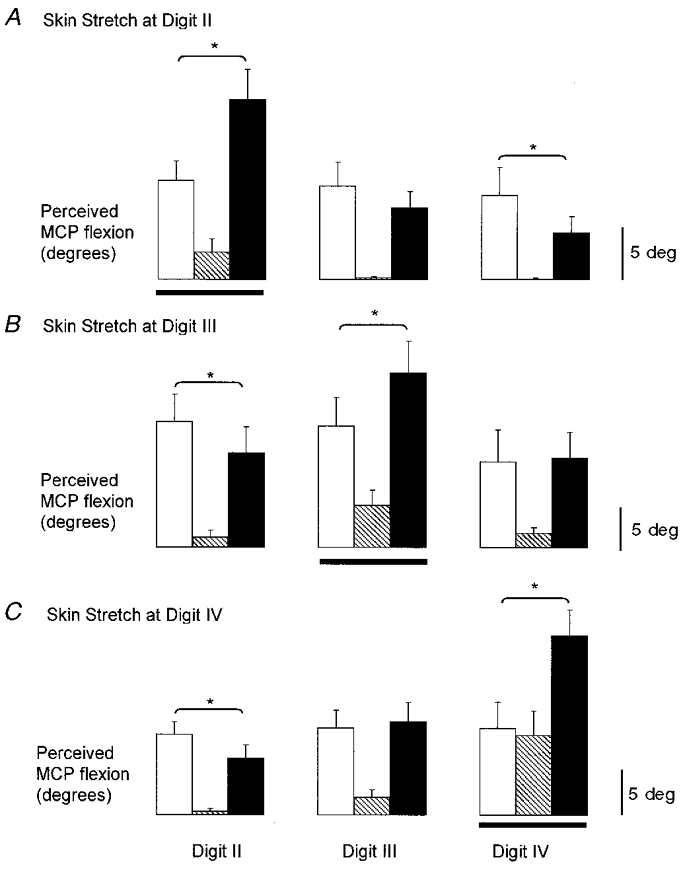
These experiments were designed to investigate illusions of movements of the fingers produced by combined feedback from muscle spindle receptors and receptors located in different regions of the skin of the hand. Vibration (100 Hz) applied in cyclic bursts (4 s 'on', 4 s 'off') over the tendons of the finger extensors of the right wrist produced illusions of flexion-extension of the fingers. Cutaneous receptors were activated by local skin stretch and electrical stimulation. Illusory movements at the metacarpophalangeal (MCP) joints were measured from voluntary matching movements made with the left hand. Localised stretch of the dorsal skin over specific MCP joints altered vibration-induced illusions in 8/10 subjects. For the group, this combined stimulation produced movement illusions at MCP joints under, adjacent to, and two joints away from the stretched region of skin that were 176 +/- 33, 122 +/- 9 and 67 +/- 11 % of the size of those from vibration alone, respectively. Innocuous electrical stimulation over the same skin regions, but not at the digit tips, also 'focused' the sensation of movement to the stimulated digit. Stretch of the dorsal skin and compression of the ventral skin around one MCP joint altered the vibration-induced illusions in all subjects. The illusions became more focused, being 295 +/- 57, 116 +/- 18 and 65 +/- 7 % of the corresponding vibration-induced illusions at MCP joints that were under, adjacent to, and two joints away from the stimulated regions of skin, respectively. These results show that feedback from cutaneous and muscle spindle receptors is continuously integrated for the perception of finger movements. The contribution from the skin was not simply a general facilitation of sensations produced by muscle receptors but, when the appropriate regions of skin were stimulated, movement illusions were focused to the joint under the stimulated skin. One role for cutaneous feedback from the hand may be to help identify which finger joint is moving.


Figures

Figure 1
Figure 1. Experimental protocol
A, method used to apply the stimuli to the stationary right hand and to monitor the resulting perceived movements with matching movements of the left hand. B, method used to stretch the skin on the dorsal and ventral sides of the MCP joint of digit II. The same technique was used at digits III and IV (not shown). C, schematic representation of the timing of stimulus delivery within an experimental block. The order of presentation of the three different types of stimulation was randomised.
Figure 2
Figure 2. Amplitudes of illusory movements evoked when skin stretch was applied over the dorsal aspect of the MCP joint of digit IV in one subject
A, raw data from a single subject when skin stretch was applied over the dorsum of the MCP joint of digit IV. The top two traces show the time course of the application of the vibration to the finger extensor tendons at the wrist and the skin stretch over the MCP joint. The bottom three traces show the resulting illusory movements at the MCP joints as indicated by the voluntary movements of the matching hand. The digit at which the skin stimulation was applied is indicated with a double underline. Flexion at the MCP joint is shown as a downward deflection. B, mean amplitude (±s.e.m.) of the illusory movements from all cycles of each type of stimulation from the data shown in A (n = 10 for each mean). Data shown for vibration as open bars, skin stretch as hatched bars, and vibration combined with skin stretch as filled bars. The skin stimulation was applied at digit IV as indicated by the horizontal line. *Significant differences between vibration and the combined stimulation.
Figure 5
Figure 5. Summary of the effects of each type of skin stimulation
Grouped data for illusory movement amplitudes evoked by combined skin stimulation and vibration over the finger extensors, normalised to the corresponding amplitudes evoked by vibration alone. Data are grouped according to the proximity to the region of stimulated skin. Data are shown for skin stretch on the dorsal side of the MCP joint (•), skin stretch on the dorsal and ventral sides of the hand (⋄), electrical stimulation on the dorsal side of the MCP joint (▵), and electrical stimulation at the distal phalanx (▪).
Figure 3
Figure 3. Amplitudes of illusory movements evoked when the skin stretch was applied over the dorsal aspect of the hand and the dorsal and ventral sides of the MCP joint of digit IV in one subject
A, raw data from a single subject when the skin was stretched on the dorsal side of the hand and compressed on the ventral side of the MCP joint at digit IV. The top two traces show the time course of the application of the vibration and the skin stretch. The bottom three traces show the resulting illusory movements at the MCP joints as indicated by the voluntary movements of the matching hand. Flexion at the MCP joint is shown as a downward deflection. The digit at which the skin stimulation was applied is indicated with the double underline. B, mean amplitude (±s.e.m.) of the illusory movements from all cycles of each type of stimulation from the data shown in A (n = 10 for each mean). Data shown for vibration only (open bars), skin stretch only (hatched bars), and vibration combined with skin stretch (filled bars). The skin stimulation was applied at digit IV as indicated by the horizontal line. *Significant differences.
Figure 4
Figure 4. Effect of dorsal and ventral skin stimulation at each digit across subjects
Mean amplitude of illusory movements from all cycles of stimulation during blocks in which the skin stretch to dorsal and ventral skin was applied at digit II (A), digit III (B) and digit IV (C). Horizontal lines indicate the digit at which the skin stretch was applied. Data shown for vibration only (open bars), skin stretch only (hatched bars), and vibration combined with skin stretch (filled bars). *Significant differences (n = 10 subjects for each mean).

References

    1. Bianconi R, Van Der Meulen JP. The response to vibration of the end organs of mammalian muscle spindles. Journal of Neurophysiology. 1963;26:177–190. - PubMed
    1. Biggs J, Horch K, Clark FJ. Extrinsic muscles of the hand signal fingertip location more precisely than they signal the angles of individual finger joints. Experimental Brain Research. 1999;125:221–230. - PubMed
    1. Brown MC, Engberg I, Matthews PBC. The relative sensitivity to vibration of muscle receptors of the cat. The Journal of Physiology. 1967;192:773–800. - PMC - PubMed
    1. Burke D, Gandevia SC, Macefield G. Responses to passive movement of receptors in joint, skin and muscle of the human hand. The Journal of Physiology. 1988;402:347–361. - PMC - PubMed
    1. Burke D, Hagbarth KE, Lofstedt L, Wallin BG. The responses of human muscle spindle endings to vibration of non-contracting muscles. The Journal of Physiology. 1976;261:673–693. - PMC - PubMed

Publication types

LinkOut - more resources

Cite

AltStyle によって変換されたページ (->オリジナル) /